May 13, 2008 - By Amy Adams
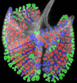
The mouse lung airway forms branches in three patterns, as illlustrated in colors: blue is domain ‘bottle-brush' branching; green is flat branching; red is 3-D right-angle branching.
Five hundred years after Leonardo da Vinci's intricate drawings of the lung drew the attention of biologists, mathematicians and engineers to the problem of how such a complex structure could form, researchers at the School of Medicine have published the first detailed description of how the intricate tree of airway tubes develops.
The painstaking work, published in the May 8 advance online issue of Nature, reveals just three branching patterns that account for the treelike structure of the lung's airways. Although the work was in mice, a better understanding of how lungs develop in mammals could help treat or prevent lung diseases in humans. Knowing the rules that govern how the branching structures form is also essential for one day regenerating several kinds of branching tissues, such as lung, pancreas or kidney, in the lab.
'We believe that setting the airway pattern is essential to patterning all the other tissues in the lung,' said lead author Ross Metzger, MD, PhD. Metzger, who is now a faculty fellow at UC-San Francisco, did the work as a PhD student working with senior author Mark Krasnow, MD, PhD, professor and chair of biochemistry at Stanford.
Figuring out how the branches form involved long hours staring at individual mouse lungs frozen in development, searching for newly forming buds and analyzing where they were positioned in relation to the main branches. All told, Metzger documented 5,051 newly forming buds, which form a remarkably similar branching pattern in each lung.
After spending hours behind the microscope, Metzger noticed some consistent patterns. In one pattern, which he called domain branching, the new branches shot out like bristles on a kitchen bottle brush as the main tube grew longer. Those bristles emerged in four neat rows at right angles to each other, forming a '+' in cross section.
He called the next pattern orthogonal bifurcation. In that pattern, the tip of a growing branch splits into left and right branches, then each of those split again into top and bottom branches. In cross section, the tips of those resulting four tubes form the corners of a square.
In the final pattern, which he called planar bifurcation, the initial growing tip divides into left and right branches, then each of those tips divides again into a left and a right branch. Each of those four resulting branches lay on the same plane as if they were drawn on a piece of paper.
Metzger said the fact that these three consistent patterns emerged is a surprise in the field. 'That was completely unexpected in the sense that the bias in the field has been that the pattern is random or just a matter of one simple branching mechanism. Somehow people weren't thinking about generating patterns,' he said.
'Scientists going all the way back to Aristotle have thought the branching structures found in many organs of the body were remarkable,' Metzger said. Most notable of those historical figures was da Vinci, who drew detailed illustrations of the lung's elaborately branching airways dated to 1508. Before Metzger's work, Krasnow, who is also a Howard Hughes Medical Institute investigator, had described many of the molecules involved in guiding airway development in flies, but nobody had done a thorough analysis of how the branches form in any mammalian organ.
In addition to describing the playbook of how branching takes place, Metzger noticed that the newly forming mouse lungs use those patterns in just three combinations. 'What's appealing about what we found is that it makes such good sense,' Metzger said. With only three patterns to create, the body can build the complex airway pattern with its large number of branches using minimal genetic information.
Metzger's next step is to find the genes and molecules that control how and when the developing lung would use one branching pattern over another.
The work was funded by grants from the National Institutes of Health.
-
 Amy AdamsAmy Adams is the director of interdisciplinary life sciences communications for Stanford University.
About Stanford Medicine
Stanford Medicine is an integrated academic health system comprising the Stanford School of Medicine and adult and pediatric health care delivery systems. Together, they harness the full potential of biomedicine through collaborative research, education and clinical care for patients. For more information, please visit med.stanford.edu.

