Stanford Fiber Tractography Lab - Anatomical Study
The brain is the seat of the central nervous system. It controls everything from clear-cut actions such as limb movement, to abstract faculties such as consciousness. Tractography allows researchers and clinicians to study living white matter anatomy over large numbers of subjects. Importantly, this represents a major breakthrough for neuroanatomists as previously, knowledge was gathered mainly from either clinical cases or post-mortem specimens which necessitated freezing and preservation prior to dissection. Aside from the damage that preparing post-mortem specimens may cause to the brain, the process of dissecting the brain from grey-to-white matter destroys the specimen and means the true connectivity of the underlying white matter cannot be accurately determined. Pioneering neuroanatomists included the likes of Broca, Wernicke, Dejerine and later on Klingler, who developed a method of preparing post-mortem tissue specimens for dissection.

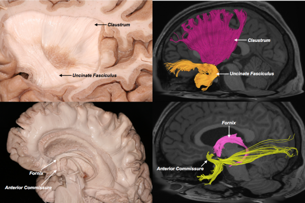
Tractography dispenses with the hurdles of acquiring, preserving and dissecting post-mortem specimens. It permits the visualization of white matter pathways by determining the diffusion direction of water molecules along axons (the brains “wiring”). This permits not only the un-altered architecture of the brain to be visualized, but also the study of white matter connectivity and calculation of differences between the left and right sides of the brain.
We have pioneered the use of tractography in neuroanatomical studies and have published in many leading neuroscience journals including Brain Structure and Function, Cerebral Cortex, Frontiers in Neuroanatomy, Neurosurgery, Journal of Neurosurgery, Neuro-Oncology and Neuroimage amongst others. Furthermore, we have pioneered the use of large-volume atlases of “averaged” data from over 1000 healthy subjects. We have also pioneered novel techniques to quantify white matter connectivity, validate tractography findings and study inter-hemispheric differences in white matter morphology.

Selected References
1. Fernández-Miranda, J. C., Rhoton Jr, A. L., Álvarez-Linera, J., Kakizawa, Y., Choi, C., & de Oliveira, E. P. (2008). Three‐dimensional microsurgical and tractographic anatomy of the white matter of the human brain. Neurosurgery, 62(suppl_3), SHC989-SHC1028.
2. Fernandez-Miranda, J. C., Pathak, S., Engh, J., Jarbo, K., Verstynen, T., Yeh, F. C., ... & Friedlander, R. (2012). High-definition fiber tractography of the human brain: neuroanatomical validation and neurosurgical applications. Neurosurgery, 71(2), 430-453.
3. Meola, A., Comert, A., Yeh, F. C., Sivakanthan, S., & Fernandez-Miranda, J. C. (2016). The nondecussating pathway of the dentatorubrothalamic tract in humans: human connectome-based tractographic study and microdissection validation. Journal of neurosurgery, 124(5), 1406-1412.
4. Meola, A., Yeh, F. C., Fellows-Mayle, W., Weed, J., & Fernandez-Miranda, J. C. (2016). Human connectome-based tractographic atlas of the brainstem connections and surgical approaches. Neurosurgery, 79(3), 437-455.
5. Meola, A., Comert, A., Yeh, F. C., Stefaneanu, L., & Fernandez‐Miranda, J. C. (2015). The controversial existence of the human superior fronto‐occipital fasciculus: Connectome‐based tractographic study with microdissection validation. Human brain mapping, 36(12), 4964-4971.
6. Yeh, F. C., & Tseng, W. Y. I. (2011). NTU-90: a high angular resolution brain atlas constructed by q-space diffeomorphic reconstruction. Neuroimage, 58(1), 91-99.
7. Yeh, F. C., Badre, D., & Verstynen, T. (2016). Connectometry: a statistical approach harnessing the analytical potential of the local connectome. Neuroimage, 125, 162-171.
8. Yeh, F. C., Panesar, S., Fernandes, D., Meola, A., Yoshino, M., Fernandez-Miranda, J. C., ... & Verstynen, T. (2018). Population-averaged atlas of the macroscale human structural connectome and its network topology. NeuroImage, 178, 57-68.
9. Yoshino, M., Abhinav, K., Yeh, F. C., Panesar, S., Fernandes, D., Pathak, S., ... & Fernandez-Miranda, J. C. (2016). Visualization of cranial nerves using high-definition fiber tractography. Neurosurgery, 79(1), 146-165.
10. Panesar, S. S., Yeh, F. C., Jacquesson, T., Hula, W., & Fernandez-Miranda, J. C. (2018). A Quantitative Tractography Study into the Connectivity, Segmentation and Laterality of the Human Inferior Longitudinal Fasciculus. Frontiers in Neuroanatomy, 12.
11. Panesar, S. S., Yeh, F. C., Deibert, C. P., Fernandes-Cabral, D., Rowthu, V., Celtikci, P., ... & Fernández-Miranda, J. C. (2017). A diffusion spectrum imaging-based tractographic study into the anatomical subdivision and cortical connectivity of the ventral external capsule: uncinate and inferior fronto-occipital fascicles. Neuroradiology, 59(10), 971-987.
12. Fernández-Miranda, J. C., Wang, Y., Pathak, S., Stefaneau, L., Verstynen, T., & Yeh, F. C. (2015). Asymmetry, connectivity, and segmentation of the arcuate fascicle in the human brain. Brain Structure and Function, 220(3), 1665-1680.
13. Wang, X., Pathak, S., Stefaneanu, L., Yeh, F. C., Li, S., & Fernandez-Miranda, J. C. (2016). Subcomponents and connectivity of the superior longitudinal fasciculus in the human brain. Brain Structure and Function, 221(4), 2075-2092.
14. Wang, Y., Fernández-Miranda, J. C., Verstynen, T., Pathak, S., Schneider, W., & Yeh, F. C. (2012). Rethinking the role of the middle longitudinal fascicle in language and auditory pathways. Cerebral cortex, 23(10), 2347-2356.
15. Yoshino, M., Abhinav, K., Yeh, F. C., Panesar, S., Fernandes, D., Pathak, S., ... & Fernandez-Miranda, J. C. (2016). Visualization of cranial nerves using high-definition fiber tractography. Neurosurgery, 79(1), 146-165.