Recent Publications
The following are general descriptions of recent publications from McNab Lab. You may also view a complete list of publications from McNab Lab.
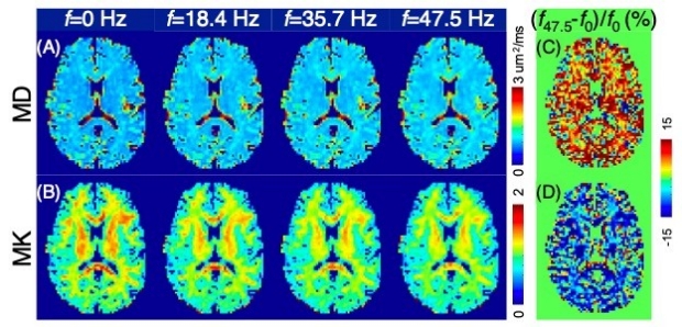
Frequency-dependent diffusion kurtosis imaging in the human brain using an oscillating gradient spin echo sequence and a high-performance head-only gradient
Description: Studying the time/frequency dependence of diffusion MRI is a promising approach to distinguish between the effects of different tissue microenvironments, such as membrane restriction, tissue heterogeneity, and compartmental water exchange. In this study, we measure the frequency dependence of diffusivity and kurtosis with the oscillating gradient diffusion encoding waveforms and diffusion kurtosis imaging (DKI) model in human brains in a high-performance, head-only MAGNUS gradient system, with a combination of b-values, oscillating frequencies (f), and echo time that has not been achieved in human studies before. Frequency dependence of diffusivity and kurtosis are observed in both global and local white and gray matter regions. A trend of decreasing kurtosis over frequency in the short-time limit is successfully captured for in vivo human brains. The effects of gradient nonlinearity on frequency-dependent diffusivity and kurtosis measurements are investigated and corrected. Our study further demonstrates the translatability of frequency-dependent diffusion MRI measurements to human brains, which may provide new opportunities to probe human brain microstructure in health and disease.
Citation: Dai E, Zhu A, Yang GK, Quah K, Tan ET, Fiveland E, Foo TKF, McNab JA. Frequency-dependent diffusion kurtosis imaging in the human brain using an oscillating gradient spin echo sequence and a high-performance head-only gradient. Neuroimage. 2023 Aug 14;279:120328. doi: 10.1016/j.neuroimage.2023.120328. Epub ahead of print. PMID: 37586445.
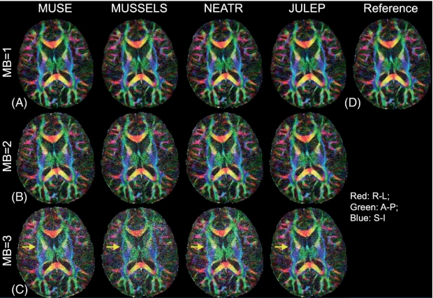
Multi-band multi-shot diffusion MRI reconstruction with joint usage of structured low-rank constraints and explicit phase mapping
Description: Diffusion MRI (dMRI) is a technique used to image the brain and other biological tissues. High-resolution images can be obtained using a method called multi-band multi-shot EPI acquisition, but it requires specific algorithms to correct phase variations between different image shots. There are two ways to correct these phase variations, and we sought to combine them to improve image quality and reconstruction efficiency. This new, combined approach is called “JULEP.” We tested JULEP on both single-band and multi-band, multi-shot diffusion data, and found that it produces better results compared to other commonly used methods, such as MUSE and NEATR. This new approach can help us obtain more accurate and detailed images of the brain, which can lead to better diagnosis and treatment of neurological conditions.
Citation: Dai E., Mani M., McNab J.A. Multi-band multi-shot diffusion MRI reconstruction with joint usage of structured low-rank constraints and explicit phase mapping. Magnetic Resonance in Medicine, 89(1):95-111, 2023.
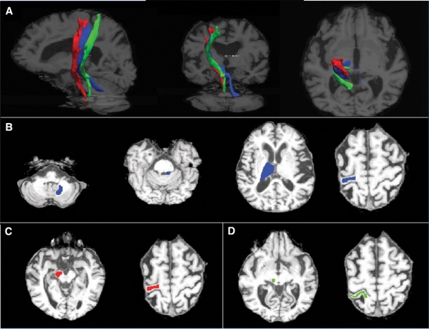
Changes in the Cerebello-Thalamo-Cortical Network After Magnetic Resonance-Guided Focused Ultrasound Thalamotomy
Description: Transcranial magnetic resonance imaging-guided focused ultrasound (tcMRgFUS) is a relatively new treatment option for movement disorders like essential tremor (ET). However, little is known about how it affects the connections within the brain. This study aimed to determine if there are microstructural changes in the purported tremor-related white matter tracts following treatment with tcMRgFUS thalamotomy. Our results showed that there were microstructural changes in the white matter tracts after treatment, and these changes were related to the location of the lesion relative to the location of the white matter tracts. These findings further verify that diffusion MRI (dMRI) tractography can play an important role in both targeting. The findings also suggest that dMRI microstructural measurements can help improve our understanding of the mechanism(s) of tcMRgFUS treatment and provide a biomarker for interpreting patient outcomes.
Citation: Thaler C., Tian Q., Wintermark M., Ghanouni P., Halpern C.H., Henderson J.M., Airan R.D., Zeineh M., Goubran M., Leuze C., Fiehler J., Butts Pauly K., McNab J.A. Changes in the Cerebello-Thalamo-Cortical Network After Magnetic Resonance-Guided Focused Ultrasound Thalamotomy. Brain Connect, 13(1):28-38, 2023.
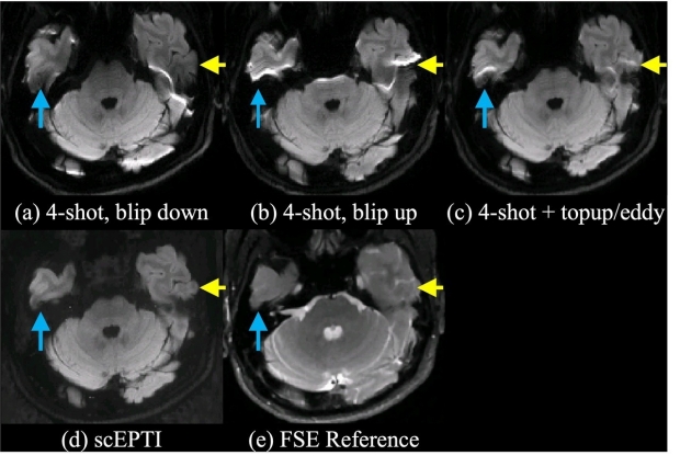
Distortion-Free Diffusion Imaging Using Self-Navigated Cartesian Echo-Planar Time Resolved Acquisition and Joint Magnitude and Phase Constrained Reconstruction
Description: Echo-planar time-resolved imaging (EPTI) is a method used to create undistorted images of body tissues. Unfortunately, it can be challenging to use EPTI with diffusion-weighted pulse sequences due to the need to correct phase variations between separate read-outs. This study developed a novel approach to data acquisition, called self-navigated Cartesian EPTI-based (scEPTI), that can correct for these variations and create undistorted images. We analyzed the different components of the EPTI signal and developed a method to match them, to further improve image accuracy. We demonstrate improvements in image quality and visualization of detailed structures afforded by EPTI. The proposed scEPTI method is a valuable tool for medical and scientific assessments of tissue microstructure.
Citation: Dai E., Lee P.K., Dong Z., Fu F., Setsompop K., McNab J.A. Distortion-Free Diffusion Imaging Using Self-Navigated Cartesian Echo-Planar Time Resolved Acquisition and Joint Magnitude and Phase Constrained Reconstruction. IEEE Transactions on Medical Imaging, 41(1):63-74, 2022.
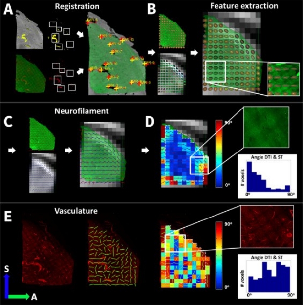
Comparison of diffusion MRI and CLARITY fiber orientation estimates in both gray and white matter regions of human and primate brain
Description: Diffusion MRI (dMRI) is a non-invasive method used to map brain microstructure. Unfortunately, this method can be difficult to interpret because it relies on indirect measures of water diffusion patterns in brain tissue. To better interpret dMRI images, researchers are comparing it with microscopy techniques that provide direct visualization of tissue components. With the advent of tissue clearing, staining and microscopy methods such as CLARITY, fibers in the brain can be visualized in 3D within an intact tissue cuboid. In this study, we compared the results of dMRI with CLARITY in the same human brain tissue. We also tested different ways of extracting information from CLARITY data to improve the accuracy of the results. This study represents an important step in improving our understanding of the brain’s structure and how different techniques can be used together to improve brain mapping.
Citation: Leuze C., Goubran M., Barakovic M., Aswendt M., Tian Q., Hsueh B., Crow A., Weber E.M.M. Steinberg G.K., Zeineh M., Plowey E.D., Daducci A., Innocenti G., Thiran J.P., Deisseroth K., McNab J.A. Comparison of diffusion MRI and CLARITY fiber orientation estimates in both gray and white matter regions of human and primate brain. NeuroImage, 228:117692, 2021.