3T3
The Richard M. Lucas Center for Imaging
The Richard M. Lucas Center for Imaging houses resources devoted to research in magnetic resonance imaging (MRI), spectroscopy (MRS) and X-Ray/CT imaging. Researchers in RSL and at the Lucas Center have pioneered MRI/MRS/X-Ray/CT technology while developing new techniques that benefit patients with stroke, brain disorders, cancer, heart disease, chronic pain, genetic disorders, joint disease and diseases of the spleen and liver. The Center supports collaborative and original research using human subjects and intact animal models.
Our facilities include three 3.0T whole-body MR systems and a 7.0T whole-body MR system, complete with patient/animal preparation facilities and image processing/readout workstations. The Axiom/Zeego Lab houses a C-arm X-Ray CT scanner that offers fluoroscopic scanning, and many other applications. A machine shop/workshop offers addition capability for miscellaneous hardware development including MRI coils and phantoms. Most of the over 100 people in RSL have office space in the Lucas Center for Imaging.
3T2 MRI Scanner

3T2 GE Premier MRI Scanner
The software and hardware currently allows the use of 128 channels at 3T2.
Capabilities supporting fMRI studies include Eprime computer, Physiologic monitoring, Eyetracker, EDA (GSR), EEG, TMS, facial camera, video projection, audio earphones, etc.
Pulse sequences include EPI, spiral, SMS and DTI.
Daily support in MR system operation and screening and safety is provided to all researchers including faculty, post-doctoral fellows, graduate students, and visiting scholars in the Lucas Center and Department of Radiology; researchers from other University departments such as Psychology, Psychiatry, Neurology, Neurosurgery, and Nephrology; and Service Center users from outside of the University.
Scanner Configuration
Software Revision: RX27.0 (upgraded December 2017)
Channels: 148
Shimming: Passive, Active & High Order
Bore Diameter: 70 cm (L-R); 49 cm vertical, no table pad; cradle width 56 cm
Head Coil Diameter: 8ch 23.5 cm; 32 ch Nova 18.4 cm; 48ch 23.5 cm
Multinuclear Spectroscopy: No
Maximum Table Weight: 550 lbs (max 550 lbs table in lowered position)
Magnetic Shielding: Active
3T3 MRI Scanner
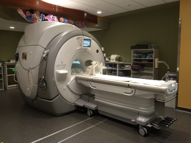
3T3 GE Discovery 750 MRI Scanner
+ InSightec Focused Ultrasound
The software and hardware currently allows the use of 32 channels at 3T3 and multinuclear spectroscopy.
Multiple options supporting fMRI studies include Eprime computer, Physiologic monitoring, Eyetracker, EDA (GSR), EEG, TMS, facial camera, video projection, audio earphones, etc.
Pulse sequences include EPI, spiral, SMS and DTI.
Daily support in MR system operation and screening and safety is provided to all researchers including faculty, post-doctoral fellows, graduate students, and visiting scholars in the Lucas Center and Department of Radiology; researchers from other University departments such as Psychology, Psychiatry, Neurology, Neurosurgery, and Nephrology; and Service Center users from outside of the University.
The InsightTec Focused Ultrasound system is used to treat human and animal models.
Scanner Configuration
Software Revision: DV26.0 (upgraded December 2017)
Channels: 32
Shimming: Passive, Active & High Order
Bore Diameter: 60 cm (L-R); 45 cm vertical, no table pad; cradle width 40 cm
Head Coil Diameter: 8ch 23.5 cm; 32 ch Nova 18.4 cm
Multinuclear Spectroscopy: Yes
Maximum Table Weight: 550 lbs (350 lbs table in lowered position)
Magnetic Shielding: Active
Lucas Mock MRI Scanner
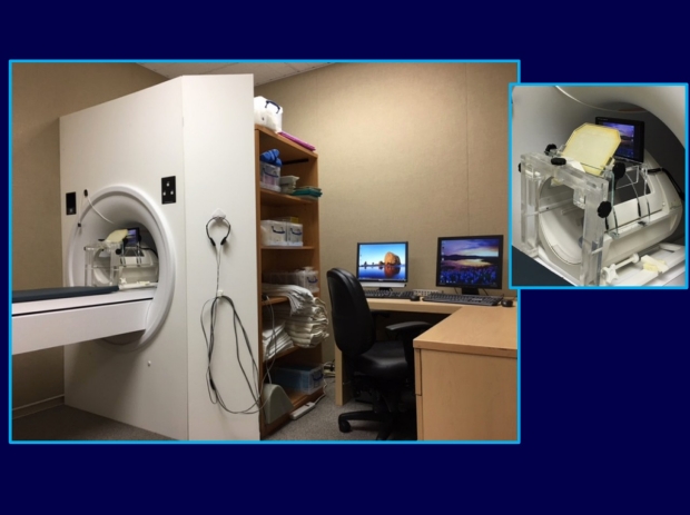
Lucas Mock MRI Scanner
Lucas Basement P020B
The MRI Simulator includes: ~60cm bore with tapered entry and flat façade with integrated control panel (Light, Fan, In/Out), lights, fan, speakers & subwoofer, motorized patient table with drag sensing safety stop, manual table release. A flat panel monitor is attached to the head coil for easy, high-resolution viewing of motion measurements, stimuli or movies for the scan subject.
MoTrak head motion tracking system that provides linear feedback of X,Y,Z movement and rotational feedback on X,Y,Z axis (Pitch, Roll, Yaw coordinate) based on user-defined thresholds.
Scanner sounds from a variety of MRI scan sequences are available including Localizer, High Order Shim, 2D T2-weighted FSE, 3D T1-weighted FSPGR, DTI, EPI, and ‘sprlio' (spiral in-out fMRI).
E-Prime® stimulus presentation software is available on a separate computer.
MRI studies require long periods of scanning with minimal participant movement. The procedure can be an unsettling experience for many participants causing excessive movement resulting in unusable data and lost funds. Special populations such as children, the elderly, and psychiatric participants, are often prone to claustrophobia and anxiety in the bore of a magnet, and consequently have a much higher rate of terminating the scan session before its completion. Some centers that have dealt with these populations estimate a 50%-80% failure rate. With the use of the MRI Simulator (also known as a mock scanner) this failure rate can often be reduced below 5%, minimizing data loss.
The MRI Simulator provides a realistic approximation of an actual MRI scanner to allow habituation and training of participants in an environment less daunting than a real scanner. The MRI Simulator introduces the participant to an authentic scanning environment, permitting them to gradually become accustomed to the scanning procedure and trained to minimize movements. Allowing the participant to acclimate to the scanner before the actual session helps to prepare them and encourages a calmer and more focused participant, ultimately resulting in more productive scanning sessions.
Focused Ultrasound (FUS) Table

InSightec FUS Table
Investigators throughout the world are currently applying MR-guided focused ultrasound (MRgFUS) for non-invasive treatments of a variety of diseases and disorders. Typically, focused ultrasound uses a large area ultrasound transducer array outside the body, focused either geometrically or electronically, to a point within the body. The amplification provided by focusing (which can be on the order of 1000-fold) provides the means to generate significant ultrasound intensities deep within the body, with insignificant ultrasound intensities in the intervening tissue. We are working with InSightec who makes a variety of systems that are integrated into General Electric MR scanners. For brain applications, InSightec has a hemisphere array.
Magnetic resonance imaging provides the means to target the ultrasound beam to the tissue of interest, monitor the therapy with MR thermometry, and assess the treatment through the variety of contrast mechanisms available with MRI. While some targeting and assessment could alternatively be done with ultrasound, MRI is the only imaging modality that provides quantitative temperature mapping over a wide range of temperatures. The key to MR thermometry is the change in hydrogen bonding with temperature, which results in a change in the electron shielding of water protons, and changes the resonant frequency by approximately -0.01 ppm/°C. In practice, temperature images are calculated from the change in phase on gradient echo images.
MRgFUS Research
Our lab is doing research in many areas of MRgFUS. Technical areas of research to support clinical applications include improved thermometry and monitoring (especially in moving organs), focal spot visualization, MR-based phase aberration correction, and workflow improvements. In addition, basic science research in ultrasound-based neuromodulation is showing that ultrasound can modulate neuronal firing. More details on our research can be found in these webpages. The future of MRgFUS holds new non-invasive treatments for a variety of diseases and disorders and also an exciting array of technical and scientific innovations.