Glioblastoma Imaging
Imaging and Grading with Multimodal Smart Probes
Gliomas comprise 80% of malignant brain tumors and can be categorized into four grades based on pathological 1characteristics such as morphological variability, angiogenesis, necrosis, and cellularity. Grade IV (glioblastoma multiforme, GBM) is the most invasive form and results in an average patient lifespan of only 15 months despite aggressive intervention. The current treatment for malignant brain gliomas involves surgical resection followed by combined radio- and chemotherapies with the extent of resection being one of the greatest prognostic indicators. Therefore, there is great interest in developing tools to provide better contrast between cancerous and normal tissues so that the maximal resection can be obtained.
Current imaging tools for glioblastomas are non-targeted and can show us where the tumor is located, but tell us little about the molecular biology behind the disease. Therefore, we aim to develop a smart probe that becomes activated upon enzymatic cleavage by furin, a serine endoprotease that is upregulated in glioblastomas. Furin is directly correlated with increased cancer aggressiveness due to its role in degrading the extracellular matrix which promotes intravasation and tumor metastasis. We want to exploit the overexpression of furin to obtain targeted, low-background tumor images and evaluate the extent of enzymatic activity in different glioma grades.
Our proposed probe uses both near-infrared fluorescence and fluorine-18 positron emission tomography imaging modalities. The probe can be used in a single- or dual-mode and could open doors to exciting preclinical and clinical applications, including: fluorescence-guided surgery, tumor grading, and evaluation of cancer-associated enzymatic processes in vivo. The modular nature of the probe is attractive as we can independently modulate the individual components to optimize spectroscopic/pharmacologic properties or exchange the peptide substrate for one of a different enzymatic target. The universality of this technology would be appealing for anyone interested in studying enzymatic activity in diseased states.

Hypoxia: Imaging Enzyme Expression in Tumors
Tumor cells exhibiting reduced oxygen concentration (hypoxia) are associated with increased aggressiveness, ability to metastasize, and resistance to therapeutic intervention. Imaging this aspect of tumor biology has clear potential to improve the staging and treatment of cancer in the clinic. Currently, nitroimidazoles labeled with 18F-fluorine (e.g., 18F-FMISO and 18F-FAZA) are the standard hypoxia-sensitive imaging probes used in PET.
For all current nitroimidazole PET imaging agents, the reactive intermediates form conjugates with nucleophilic biomolecules and these metabolites are not confined to the intracellular environment. Diffusion and circulation of these radioactive metabolites causes significant non-target tissue accumulation of radioactivity, and delay in background clearance, resulting low signal-to-noise ratios. As a result, 18F-FMISO and 18F-FAZA have limited sensitivity and specificity for hypoxia in cancer, which limits the clinical utility of these agents.
Despite over 20 years of engineering of nitroimidazole-based hypoxia imaging probes, the performance of these imaging approaches has remained largely unchanged, and the potential of this imaging modality has not been realized despite over 60 years of studies demonstrating the clinical significance of tumor hypoxia.
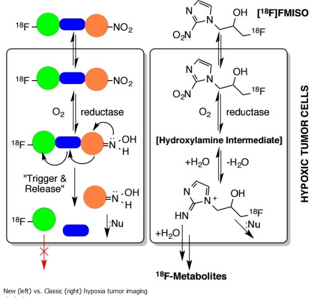
We propose an entirely novel non-nitroimidazole-based “trigger and release” strategy for imaging hypoxia with PET. This new tactical approach to imaging probe design features a reducible “trigger” as the reductase target, an irreversibly-cleavable linker to favor “release” of the masked imaging agent, and a customizable 18F-labeled moiety for intracellular retention or “trapping” under hypoxic conditions. Since the trigger is mechanistically detached from the 18F-bearing group following reduction, the fate of the modified trigger subunit is irrelevant for imaging, which detects only emissions from the 18F atom. Under normoxic conditions, the trigger is efficiently back-oxidized to the parent compound by molecular oxygen, diffuses back into the circulation and is excreted.
In vitro studies have confirmed the mechanistic release of the 18F-moiety, and since the approval of the Investigational New Drug Application by the FDA, the clinical phase of the 18F-FMISO study has commenced.