Center for Cancer Nanotechnology Excellence
for Translational Diagnostics (CCNE-TD)
Stanford Environment
I. Nanomaterial Characterization and Animal Resources
Stanford Nanotechnology Characterization Lab (NCL): Our original Nanoinformatics Core in our past phase-I Center, CCNE-TR, centralized efforts to provide data analysis and knowledge management for our CCNE members and to the NCI Alliance. Through this early effort, we were able to address the nano needs of research projects and cores by facilitating representation and delivery of nanocharacterization data to caNanoLab as well as respond to information retrieval needs of the CCNE researchers. This core was dedicated to laying the foundation of cancer nanoinformatics and to helping develop just such a resource. Overall, the special responsibility of this core was to facilitate interactions between the different NCI Alliance members, especially from a data exchange perspective.
Our current phase-II Center, CCNE-T Nanocharacterization and Nanofabrication Core, consists of two entities: 1) the Stanford Nanocharacterization Laboratory (SNL), which provides resources for analysis of micro- and nano- structures and 2) the Stanford Nanofabrication Facility (SNF), which offers resources for fabrication. This highly functional core represents a merging of Cores 1 and 2 of the original CCNE-TR program. By integrating fabrication and analysis services, this Core provides complete and essential nanotechnology and engineering support for the projects in this Center as well as performing original research associated with its mission.
Building on our efforts to also assist in defining nanoinformatics, along with building much-needed resources, our current CCNE-T, regularly sends to the NCI Nanotechnology Characterization Laboratory (NCL) nano particles that they not only study for toxicity, but also help us to better characterize. In this way, the NCI’s NCL provides a significant service that ultimately helps us with translation and FDA issues. In our current proposal, Center for Cancer Nanotechnology Excellence for Translational Diagnostics (CCNE-TD), our communication and sharing of materials will increase significantly as we move into a dedicated translational phase of our program.
Animal Toxicity and Resources: In addition to the significant animal resources available to the CCNE-TD (see section II, E-H), our group is experienced in conducting toxicity studies in small animal tumor models. Because of our longstanding expertise and resources, our practice has been to conduct early toxicity studies for newly developed compounds and submit for further study to the NCI’s Nanotechnology Characterization Lab (NCL). The NCL resources provide an independent preclinical assessment of nanomaterials submitted and in a variety of xenograft and genetically engineered mouse models that richly expand our early studies. We will continue this practice for the proposed CCNE-TD.
II. Resources - Stanford University School of Medicine
The Stanford Cancer Institute (SCI)
The Stanford Cancer Institute (SCI) focuses the world-class expertise of more than 300 researchers and clinicians on the most critical issues in cancer research and medicine today. These dedicated individuals work together in multidisciplinary teams to unravel cancer’s secrets and to transform the latest detection, diagnosis, treatment and prevention discoveries into the most advanced patient care available. Combining these advances with comprehensive support services, the SCI is committed to giving patients every clinical and technological advantage in the prevention and treatment of cancer. The SCI advances its mission through: Comprehensive patient care; Research discovery and medical innovation; advanced professional training; state-of-the-art facilities; community outreach; leadership.
Home to a closely integrated research and patient care campus, Stanford Medicine offers immense opportunities for cross-disciplinary collaboration across the bench-to-bedside continuum. Unique among medical institutions, the center situates a world-class medical school next to its primary teaching hospital while itself sitting within a major university setting. As part of the world-class research corridor running through Northern California, the center also benefits from important industry collaborations.
Opened in March 2004, the Stanford Cancer Center offers the latest advances in patient-focused treatment in a warm and supportive environment. Consolidating the medical center’s resources for diagnosing and treating cancer, the new cancer center enhances Stanford’s team approach to care while enabling patients to stay in one location for procedures and tests. Located directly across the street from the Cancer Center is the Cancer Clinical Trials Office (CCTO). The CCTO provides regulatory, administrative, research, and educational services to Cancer Institute investigators conducting clinical trials. Programs of the resource serve to increase awareness and accrual to clinical trials as well as to improve the quality and efficiencies of conducting clinical trials in compliance with regulatory, documentation, and oversight requirements. Many departments in the School of Medicine, including the Department of Radiology, partner with the CCTO to provide rich support services for patients, their families, and clinicians; to centralize and standardize data collection and clinical cancer research reporting, to develop outreach programs; and to promote interdisciplinary collaborations and translational medical research. All of our clinical collaborators are members of the Stanford Cancer Center and the Cancer Institute.
Stanford University is generally considered one of the most highly regarded centers for biomedical research worldwide. With 1760 faculty members, including 400 tenure-line faculty and >1000 doctoral students in the School of Medicine, 18 Nobel Laureates and more than 100 members of the National Academy of Sciences and Institute of Medicine, Stanford University has a diverse and immense scope of high quality research activity. The Department of Radiology at Stanford University is recognized as one of the top radiology training and research programs in the world.
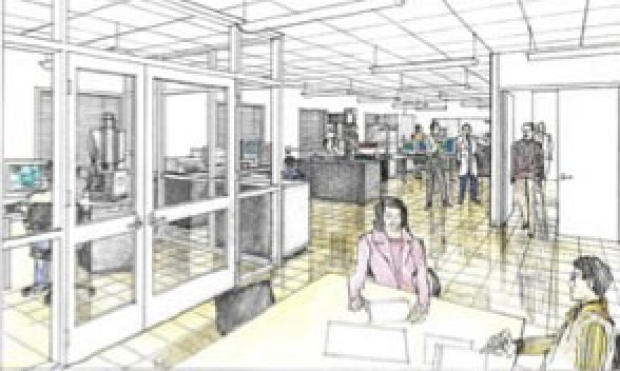
Stanford University has strongly believed that its strengths in the School of Medicine, Engineering, and Humanities & Sciences need to be leveraged towards advancing biomedical sciences. For this reason, a key effort known as the Bio-X Program was established and a new building of 225,000 square feet, known as the James H. Clark Center, was constructed. This was an investment of over $175 million in construction costs and infrastructure that brings together scientists from many disciplines including Chemistry, Engineering, Developmental Biology, Molecular Pharmacology, Medicine, Surgery, and Radiology. All the Bio-X Program Scientists share a common goal to advance Biomedical Science. The proposal will benefit tremendously from the environment at Stanford, The Schools of Medicine, Engineering and Humanities & Sciences and the Bio-X that fosters highly multidisciplinary efforts including nanotechnology research.
The Department of Radiology in the School of Medicine
For more than one hundred years, Stanford’s Department of Radiology has been making contributions to medical and surgical advances by pioneering innovations in image-based research, patient care, and education. Researchers in the Radiology Department have initiated more than 100 patents over the past 5 years. Their work has enabled Stanford Radiology to be among the world’s leaders in creating platforms for Molecular Imaging, including nanotechnology applications, CT and MRI technologies. Among U.S. medical schools, Stanford Radiology now ranks as the third highest NIH-funded Radiology Department and has five NIH-funded Centers of Excellence, the National Center for Advanced Magnetic Resonance Technology (CAMRT); the in Vivo Cellular and Molecular Imaging Center at Stanford (ICMIC); the Center for Cancer Nanotechnology Excellence and Translation (CCNE-T); the Magnetic Resonance Imaging-Guided Cancer Interventions (MRgCI), and the Center for Cancer Systems Biology (CCSB). All but one of our research facilities are within 5-10 minutes walking distance from each other, including the School of Engineering and the Stanford Hospital, thereby providing a unique concentration of a large variety of high quality resources and scientists that enable interdisciplinary basic and translational research. The Canary Center at Stanford for Cancer Early Detection is located about a short drive or bike ride away (1.5miles) on Porter Drive at the Technology and Innovation Park (see below).
The Canary Center at Stanford for Cancer Early Detection
The Canary Center for Cancer Early Detection was established in 2009 as a partnership between the Canary Foundation and the Stanford University School of Medicine. The Foundation contributed $15M to the $5M that the Department of Radiology donated to jumpstart the Center, recruit faculty and staff, and buy equipment. Stanford University also committed 8 new faculty billets (FTE’s) for the growth of the molecular imaging program and for early cancer detection efforts. The Center is the beneficiary of the highly productive collaborative network established by the Canary Foundation, as well as ongoing financial and technological support from the Foundation on multiple levels. Ongoing Foundation fundraising activities continue to provide the Center with infrastructure, seed grants, and industry partnerships that bring services and technology platforms to the Center. The Canary Foundation also employs a highly effective team of program managers who are devoted to the management and coordination of collaborative research ventures, channeling resources and serving as liaisons to the Center.
The Canary Center has recently moved to the Stanford Technology and Innovation Park where the Department of Radiology occupies 55,000 square feet of newly renovated office and laboratory space. The newly- renovated research facility includes a suite of molecular and cellular biology labs, office space, and shared resource core laboratory facilities for proteomics, bioinformatics, chemistry, and a biospecimen repository. The Proteomics Core Facility is designed to accommodate numerous technologies, including several mass spectrometry platforms dedicated to the discovery, verification, and validation of blood biomarkers and imaging targets for early cancer detection. The Chemistry Core Facility is dedicated to the development of molecular imaging agents for multiple modalities, including positron emission tomography, ultrasound, magnetic resonance imaging, photoacoustic, and optical imaging. The Bioinformatics Core Facility is devoted to developing solutions for collaborative translational research, including the implementation of software platforms for specimen tracking, laboratory informatics systems, and cross-platform data analysis, sharing and storage. The Cell and Molecular Biology Core Facility is equipped with state-of-the-art shared resources for cell culture, microscopy, and molecular biology. The Small Animal Imaging Core Facility is designed to accommodate rodent imaging and currently houses PET optical imaging platforms. See the Equipment section for the tremendous array of resources located at the Canary Center.
The Lucas Center and Radiological Sciences Laboratory (RSL)
The Richard M. Lucas Center for Imaging was established in 1992 and is the primary, centralized resource dedicated to biomedical imaging on the Stanford University campus. The original Lucas Center quickly outgrew its space and was expanded to accommodate an increased number of faculty and postdoctoral students in 1997. In 2005, this space was expanded yet again, adding 20,000 nasf to the existing 17,000 nasf. Approximately 90% of the new 20,000 nasf is dedicated to molecular imaging and nanotechnology. This expansion presented an exceptional opportunity for the Radiological Sciences Laboratory (RSL) and Molecular Imaging Program at Stanford (MIPS) to expand and build on existing research programs. This facility is located on the Stanford campus one block from the School of Medicine and Stanford Hospital. Major imaging devices available for research studies include three 3T and one 7T MR imagers, and one digital x-ray fluoroscopic/angiographic system.
With its 37,000 nasf of space dedicated to molecular imaging, nanotechnology, MRI, CT, PET, PET/CT, and spectroscopy research, the Lucas Center is one of the few centers in the world with major centralized resources devoted to research in the radiological sciences where both basic and clinical scientists are housed. The Lucas Center builds on a long-standing and very close working relationship between faculty and students of RSL, MIPS, and members of Bioengineering, Electrical Engineering, Psychology, Psychiatry, Neurobiology, and other departments. The Center provides office and laboratory facilities for over 10 full-time faculty and their complement of more than 75 postdoctoral fellows, students, and staff. With these resources, the Lucas Center is well suited for MR scanning of patients, animals, and normal volunteers comfortably and safely. Examples of labs and resources
The Radiological Sciences Laboratory (RSL), co-directed by Drs. Gary H. Glover and Kim Butts Pauly, is a section within the Stanford University Department of Radiology, and is located in the Lucas Center and the Porter Drive facility. The RSL is comprised of 10 full-time basic science faculty, averages 50 predoctoral and postdoctoral students, and hosts numerous other fellows and visiting scholars. The mission of the RSL is to support basic research in imaging science, provide a nurturing environment for students and postdocs, and collaborate with others within and outside of the Department. The following programs and labs fall under the umbrella of the Radiological Sciences Laboratory.
The Magnetic Resonance Laboratory (SMRL, smrl.stanford.edu) provides nuclear magnetic resonance (NMR) spectroscopy, instrumentation, and expertise. The Laboratory’s primary focus is on the spectroscopy of bio-molecules, including studies of polymers, minerals, soils, and extracts. Laboratory equipment and services include: 800MHz Varian Inova Spectrometer, 600MHz Varian Inova Spectrometer, 500MHz Varian Inova Spectrometer, and staff assistance/consultation.
The Stanford High Field Program, led by Dr. Brian Rutt, aims to develop and make available to the research community at Stanford a next-generation 7 Tesla whole-body magnetic resonance imaging (MRI) system, specifically the GE Discover MR950 7.0T system with parallel transmit capabilities, to serve as a platform for cutting-edge imaging technology research and development, as well as for radiological and neuroscience research. The high field program at Stanford is interdisciplinary, bringing together researchers from the specialties of physics, engineering, bioengineering, biology, physiology, radiology, neurology, psychiatry, and psychology. The whole-body 7T MR facility housed at the Lucas Center will act as a catalyst and common platform for this group to create, refine, implement, validate and utilize the most advanced forms of magnetic resonance imaging. Major patient-based imaging research applications of the next-generation 7T MRI platform include studies of brain development, psychopathology, drug dependence, alcohol-induced brain damage and its functional consequences, neurodegenerative processes, Williams, Turner and fragile X syndromes, brain injury, breast cancer, joint injuries, and therapeutic interventions associated with some or all of the above. Major technology development directions that will be enabled by this next-generation 7T MRI platform include MR spectroscopic imaging (MRSI) of the proton (1H) nucleus as well as non-proton nuclei, in both brain and musculoskeletal systems, advanced perfusion and diffusion tensor imaging in brain, whole breast imaging, and, importantly, parallel transmit technology for mitigating B1 inhomogeneities that limit the use of high magnetic field MRI in any organ system. Our objective is to develop software and hardware methods to allow 7T MRI to have a much greater impact on clinical research than possible before, as well as to extend the capabilities of high-field MRI to unprecedented levels of spatial resolution, metabolite and iron sensitivity, and tissue characterization. Projects within the high field MR program are highly compatible with the mission of the Department of Health and Human Services and relevant to public health. The proposed research will take place at interdisciplinary laboratories directed by international leaders in imaging research: high field and high sensitivity MRI methodology development (Dr. Brian Rutt, PI), developmental disorders and clinical neuroscience (Dr. Allan Reiss), DTI methodology development (Dr. Roland Bammer), musculoskeletal disorders and radiological research (Dr. Garry Gold), breast MRI methodology development (Dr. Brian Hargreaves), parallel transmit and RF pulse technology development (Dr. John Pauly), psychiatric disorders and neuroimaging (Drs. Adolf Pfefferbaum and Edith Sullivan), MR spectroscopic imaging methodology development (Dr. Dan Spielman), psychiatric disorders and clinical neuroscience (Dr. Edith Sullivan), cognitive neuroscience and neuroimaging (Dr. Brian Wandell) and neurovascular imaging (Dr. Greg Zaharchuk).
The Advanced Imaging Techniques Laboratory, directed by Dr. Rebecca Fahrig is located in the Grant Building of the School of Medicine, immediately contiguous with Stanford University Hospital. It contains two X-ray laboratories with a combined space of approximately 1000 sq. ft:
The Axiom Laboratory is a fully featured clinical-grade Zeego angiography suite, from Siemens Medical Solutions, where minimally invasive experimental procedures are performed by clinicians on animal models for developing new clinical techniques. The system consists of a ceiling mounted rotational C-arm X-ray system with a-Si large area flat panel digital detector (158 micron pixel size), which has a high dynamic range. The system can be used in standard fluoroscopic mode or in a rotational-CT mode creating 3D volumes from angular projections acquired through a 220 degree rotation. The projection data can be acquired in as little as 5 seconds, or as many as 500 projections can be acquired in 20 seconds. Complete CT volumes with isotropic pixel sizes can be reconstructed down to 200 microns. Contrast enhancement and DSA are performed using a MEDRAD programmable contrast injector. The Zeego system also includes two X-Leonardo workstations for image volume reconstruction and rendering and a large DICOM server, each with fast dual Xeon processors and loaded with Siemens CT and volume render software. The suite also provides a test bed for a number of Stanford – Siemens collaborations.
The Digital X-Ray Imaging Laboratory features a bench top laboratory system setting for cone-beam CT development. The system, mounted on an optical bench, features a Varian G1590SP X-ray tube (0.3/0.6 mm focal spots) powered by a CPI 100 kW R/F X-ray generator and a Varian a-Si digital X-ray detector that can run in R & F and pulsed fluoroscopic modes. The 30 x 40 cm flat panel detector has a pixel size of 194 microns that yield 16-bit data at various frame rates up to 30 FPS, and includes programmable dynamic and dual gain corrections. Sub-portions of the panel can acquire at even higher data rates. System components are mounted on a 9-axis motion control system that can manipulate test objects at the imaging axis center of rotation, as well as X-ray source and detector with micron precision and repeatability. System motion, X-ray triggering, and detector image acquisition are performed through coordinated programmable computer control. High-end workstations featuring fast dual Xeon processors are employed for projection image acquisition and CT-volume reconstruction. Test objects in the lab include an inventory of relevant anthropomorphic phantoms including chest phantoms featuring various interchangeable lung and heart inserts. The lab features test and measurement equipment for electronics, dose calculation, X-ray physics, and image quality. The lab also provides desk space for research development and data analysis.

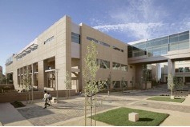
James H. Clark Center
The James H. Clark Center, the hub for the Bio-X program, fosters an unprecedented degree of collaboration between scientists from different disciplines in order to meet some of the most pressing scientific and medical challenges of the coming decades. Such challenges can no longer be met by individual disciplines working in isolation, but require the combined expertise of multi-disciplinary teams. The Clark Center lies at the heart of the Stanford campus between the core campus science engineering buildings and the hospital and medical facilities. Located on primary routes between the campus and the medical center, the building acts as a social magnet encouraging chance encounters and informal meetings between lecturers, researchers and students from diverse academic backgrounds. The lab interiors are a dramatic departure from tradition. The building has been turned inside out, with 'corridors' replaced by external balconies, enabling completely flexible lab layouts. The three-story building takes the form of three wings of laboratories centered on an open courtyard overlooked by balconies. The Clark Center is home to approximately one half of the Radiology 3D Imaging Laboratory, the Molecular Imaging Lab, and the small animal imaging, which offers a full complement of small animal imaging and surgical devices, one floor below Dr. Sam Gambhir’s Molecular imaging Laboratory. This facility offers state-of-the-art imaging using optical imaging, microscopy, CT, SPECT/CT, a 7T MRI as well as micro and macrotome facilities. The Clark Center, in addition to the Lucas Center, houses significant resources supporting the Molecular Imaging Program at Stanford (MIPS).
Molecular Imaging Program at Stanford (MIPS)
The Molecular Imaging Program at Stanford (MIPS), directed by Dr. Sam Gambhir, is a section within the Stanford University Department of Radiology with labs located in the Clark Center, the Alway Building, the Edwards Building, the Lucas Center, and the Porter Drive facility. Approximately 10,000 nasf of the Lucas facility is used for radiochemistry and labeling procedures. The following labs and programs present examples of resources that fall under the umbrella of the MIPS program. The Clark Center that follows also houses MIPS resources, such as the Small Animal Imaging Lab described below.
The Stanford Radiology Cyclotron, led by Dr. Frederick Chin, housed in the Radiochemistry Facility (RF), develops and offers radiotracers for early detection and therapeutic monitoring of disease in both preclinical and clinical imaging settings. The faculty, staff, and postdocs numbers around 30 people. Instruments include two GE radiosynthesis modules (TRACERlab FX-N Pro and FX-FN), two Agilent HPLC with autosampler systems, and a prototype miniature microwave system (CEM PETwave). A clinical radiochemistry laboratory is in the final stages of completion in the new Nuclear Medicine and Molecular Imaging Clinic, which opened in October 2010. This extra lab space provides clinical-grade radiopharmaceuticals to meet essential clinical radiochemistry demands while abiding by current regulatory policies. The existing radiochemistry labs continue to provide tracers for pre-clinical investigations and maintain [C-11] carbon dioxide and [F-18] fluorine gas radiochemistries. The facility provides routine clinical tracers for use at the Stanford Hospital. Fluorine-18 labeled fluorodeoxy-glucose (FDG) is produced daily (6-days/week) and is made using the new FASTlab FDG system with much higher yields relative to the MX-FDG module. Nitrogen-13 ammonia (myocardial perfusion assessment) and fluorine-18 sodium fluoride (bone imaging) are also synthesized for the clinic as needed. GE TRACERlab modules are the workhorses in the lab and perform the syntheses of 18F and 11C-labeled radiotracers for many collaborations at Stanford and pharma. These modules enable performance of new radiochemistries such as [18F] FSPG (imaging cystine/glutamate exchanging in tumors) for human studies. Additional PET radiotracers that study the mechanisms and treatment of cancer as well as neurological disorders will soon become available to meet the increasing needs for performing preclinical ([11C]raclopride, [18F]saxitoxin, [18F]FBR, [18F] FTC-146, [18F]FA-YF3) and clinical ([64Cu]rituximab, [18F]Avid/ Bayer/GE compounds, [18F]FSPA-RQ, [18F]AraG) research studies with PET.
The Clark Center three Radiology labs, including a small animal imaging laboratory, molecular imaging laboratory, and a full complement of small animal imaging and surgical devices one floor below Dr. Gambhir’s molecular imaging lab. This facility offers state-of-the-art imaging and includes the following resources.
Animal Imaging Resources
MicroPET Imaging System (Concorde, R4): Concorde Microsystems Rodent 4 ring MicroPET tomograph, capable of acquiring list mode data and sorting the results to arbitrary (sub second) frame durations. Absolute system sensitivity is ~2.3% at the CFOV and spatial resolution is ~2 mm isotropic. Maximum A Posteriori, Ordered Subsets and Filtered Back-projection image reconstruction are available. The system has 20 cm transverse field of view (FOV), and 8 cm axial FOV. The Concorde R4 MicroPET™ tomograph includes a rotating point source that spirals across the field of view (FOV). The location of the source is tracked with the aid of a laser light and a motion encoder and is inserted in the list-mode data stream. The firmware and software of the system allow windowing the detected events for the acquisition of transmission scans in either singles or coincidence mode. Typical pre-injection singles transmission scans require 20 minutes for two complete passes of the point source from every location of the FOV. In the same fashion, blank calibration acquisitions can be acquired and result transmission images that can be used for attenuation correction. Located in the Clark Small Animal Imaging Facility.
In Vivo Optical Imaging System (Calipers Inc.): This optical imaging system consists of a light-tight box with a mounted cooled charge couple device (CCD) camera (IVIS™). The system is fully calibrated using a standard "hockey-puck" with scintillation cocktails with four small point sources of light. Standard software routines are provided with the system and bioluminescent signal can be quantified in photons/sec/cm2/ steridian. Three other IVIS™ systems with fluorescence adaptors are also available. There is significant in house expertise to modify these systems with different filters, light sources and CCD cameras as needed. Located in the Clark Small Animal Imaging Facility.
Small Animal High-Resolution Ultrasound (VisualSonics, Vevo): This high-resolution ultrasound imaging system enables in vivo visualization, assessment, and measurement of anatomical structures and hemodynamic function in longitudinal imaging of small animals. Real-time images can be acquired in B-mode, Pulsed-wave Doppler Mode, and M-mode at a frame rate of 30Hz. Real-time microvisualization (RMV) scanheads of different centering frequencies (40 MHz, 30 MHz, 25 MHz) can be used to optimally image structures at resolutions down to 30 microns or guide injection of genes or cells into mouse embryo, mouse heart, and rat heart, respectively. The built-in Vevo Software allows myriads of measurements including placental diameter and depth, left ventricular volumes, and velocity time integral across all major valves. A Vevo Integrated Rail System is also provided to allow quick alignment and adjustment of RMV probe and the associated injection system. Located in the Clark Small Animal Imaging Facility.
Small animal High-Resolution Computer Tomography (Explorer RS Micro CT System): This high-resolution microCT system allows acquisition of 3-D images of both in vivo and in vitro specimens at 45 or 90 μM for whole body small animals and in vitro specimens at 27 µM. This system use volumetric Conebeam CT technology that allows the entire volume of a sample to be imaged in one rotation and provides exceptional image quality with short scan times with great signal to noise ratios. Located in the Clark Small Animal Imaging Facility.
Digital Whole-Body Autoradiography (DWBA) Facility: The autoradiography facility includes both a Leica cryostat for whole-body sectioning of small animals and a Molecular Dynamics phosphor imaging system, which produces autoradiograms with a resolution as high as 50 microns. Mice injected with a tracer are sacrificed, frozen in carboxy-methyl-cellulose and whole-body sectioned at 25-50 micron thickness. Tissue slices are exposed for 4-72 hours to digital plates, which are then scanned in a phosphor imager. Photographs of whole-body sections can be compared with autoradiograms to correlate anatomy and probe localization. Using radioactive standards, full quantification is possible. Animals scanned by MicroPET can subsequently be sacrificed and used for high-resolution tracer distribution analysis by DWBA. Animals scanned optically can also be sectioned for comparison of optical signal with anatomy. Located in the Clark Small Animal Imaging Facility.
Microscopy Facilities: These facilities are in the Beckman Center, which is within one minute from the Clark Center. Confocal Laser Scanning LSM 510 equipped with a coherent mira 900 tunable Ti: Sappire laser for two photons. Vendor- Zeiss. Features- Simultaneous collection of single and multiple excitations of different fluorophores. Filters- DAPI< FITC, Rhodamine also does DIC imaging. Location- B050, Beckman Center. Deltavison deconvolution microscope. Vendor- Applied precision Inc. Features- Two cooled CCD camera, Can image triple labelled specimen at all three wavelengths. Filters- DAPI, FITC, GFP, Rhodamine, Cy3, Texas Red, Cy5. Location -B050, Beckman Center Website: http://taltos.stanford.edu/pages/cocoa_dv.html In addition, two simpler fluorescent microscopes are available in the Clark Center. These are Zeiss and Leica systems with various filter options.
Cyclotron Facility: The Cyclotron Facility is housed in the Lucas Expansion building on the main Stanford campus. This building houses a GE PETtrace 18MeV cyclotron. The facility is managed by cyclotron chemists and a cyclotron physicist. The cyclotron control system is PC based and fully automated, requiring little operator intervention. Curie quantities of F-18 and C-11 and deci-curie amounts of N-13 isotopes are routinely realized with these cyclotrons. Availability of FDG/Fluoride ion, and availability of reporter gene imaging probes (e.g., FLT) is three times per week. The F-18 is used with automated nucleophilic synthesizers (GE) in order to synthesize FHBG using a software based automated module. We also have the capability to perform Iodinations using I-124 (e.g., for FIAU). Equipment provided at no direct cost to sponsor.
The Stanford Vivarium is housed directly next to the Clark Center. It is staffed by veterinarians and by certified laboratory animal care technicians who provide animal care and procure animals from licensed vendors. A newer small animal holding facility for serial microPET and other imaging studies has recently been constructed and is housed within the Clark Center (directly adjacent to the small animal imaging suite). This facility is also run by the same veterinarians; the facility is accredited by the American Association for the Accreditation of Laboratory Animal Care. All animal studies are required to have an approved policy number regulated by the Institutional Animal Research Committee (ARC).

Veterinary Service and Comparative Medicine Animal Resources
Comparative Medicine is a distinct discipline of experimental medicine that uses animal models of human and animal disease in translational and biomedical research. The Department of Comparative Medicine at Stanford is an academic department whose faculty teach at the undergraduate, graduate, professional and post graduate levels. The Department's faculty are also engaged in collaborative and comparative research, with animal model expertise and programs in veterinary pathology, pain and anesthesia, rodent reproductive biology, infectious disease, cancer, bioengineering and neuroscience. In addition, the veterinary faculty in the Department of Comparative Medicine have oversight responsibility for the campus-wide animal research program and provide clinical service in the Veterinary Service Center. Our mission is to advance human and animal health through outstanding research, veterinary care and training.
The concept of “One medicine, one health” is based upon a fundamental idea: advances in both human and veterinary medicine are dependent on overlapping technologies and research discoveries. This is the essence of Comparative Medicine-- the study of the close connections between human and animal health and disease.
The faculty in the Department of Comparative Medicine are basic researchers or veterinary clinician scientists, all working toward one health. Basic scientist faculty use animal models of epilepsy, neuronal reorganization and recovery after injury, and cortical neuronal circuitry to study physiological and patho-physiological processes. The veterinary clinical faculty in the Department of Comparative Medicine research interests focus on topics pertaining to laboratory animal and wildlife health, and on animal models of human disease. The species studied are diverse, ranging from rodents to the African Clawed frog.
The Veterinary Service Center serves all aspects of the Department and assures that the use of all animals is humane and complies with all relevant policies and legal requirements. The functions of the VSC include: the procurement of animals for research and teaching; the provision of veterinary care; the provision of animal husbandry services; the oversight of animal holding facilities; and the provision of special services that facilitate animal research
Translational Applications Service Center (TASC)
Translational Applications Service Center (TASC) facility in the Department of Medicine is a fee-for-service research laboratory serving the Stanford University scientific community, as well as outside investigators. It offers a variety of analytical services, technical and scientific consulting and training, as well as pay-per-use research equipment, which allow investigators shared access to technologies that support innovative research in translational medicine. By providing centralized oversight and infrastructure for conducting translational research studies, as well as access to high-quality, cost-efficient, state-of-the-art proteomic, molecular and genomic technologies, TASC's overall mission is to serve as a comprehensive resource for accelerating the pace of advances in patient therapy and diagnosis by enhancing basic research in the early stages of translation to the clinical setting.
We can provide scientific expertise and support for standard and customized assays, from the initial study design to the final interpretation and statistical analysis of experimental data. Our standard services also include clinical sample acquisition, processing and archival for conducting inter-disciplinary translational research applications.
Translational Research and Applied Medicine (TRAM)
The Translational Research and Applied Medicine (TRAM) Program is designed to focus on removing barriers and communication gaps between scientists and the clinicians within the Department of Medicine. TRAM was established to provide an infrastructure to rapidly translate novel genomic/proteomic, nanoscale and imaging research discoveries from the laboratory to the clinic and facilitate bench-to-bedside development of cellular therapies. Our initiatives include research facilities and pilot grants that support translational projects of faculty, clinical fellows and students. The program also fosters education by organizing a series of seminars, symposia and an annual retreat. Our program is interested in all phases of translational research, as well as both directions of translation (from bench to bedside and from bedside to bench) and also in the extension to the population level.
Integrative Biomedical Imaging Informatics at Stanford (IBIIS)
The Integrative Biomedical Imaging Informatics at Stanford (IBIIS) Laboratory, co-led by Drs. Napel (PD) and Plevritis, consists of a section within the Department of Radiology of radiology faculty and their students, staff, and post-doctoral associates. Its mission is to advance the clinical and basic sciences in radiology, while improving our understanding of biology and the manifestations of disease, by pioneering methods in the information sciences that integrate imaging, clinical and molecular data. This expertise available in this section spans image, information and natural language processing, informatics and bioinformatics, and computational systems and cancer biology. It is a highly collaborative group, as epitomized by their collaboration with Interventional Radiology for this and other projects.
The Stanford Radiology 3D and Quantitative Imaging (3DQ), co-directed by Drs. Dominik Fleischman, Roland Bammer, and Sandy Napel, located in both the Grant Building and the Clark Center, is dedicated to the clinical and research applications of advanced computer graphics for the visualization of medical data. The clinical arm of the laboratory, staffed by full-time laboratory managers, a software engineer, two administrative assistants, and five full time 3D technologists, processes between 900 and 1000 cases per month, generating alternative visualizations and quantitative analyses for radiologists and referring physicians. These 700 nasf facilities house a number of computer workstations, as itemized in the Major Equipment section. Software includes modules for complete 3D Analysis, Vessel Analysis, CV Flow, Mass Analysis, CT Perfusion, Smart Score, Cardiac IQ (including function), flythrough auto navigation for CT Colonography, Advanced Lung Analysis, Data Export, coronary calcium (quantified in Agatson score, volume, and mass), endovascular 3D stent planning tools, time-density analysis, lung nodule tracking & analysis, MR oximetry and post-processing for large CT angiography datasets of the extremities. The laboratory also hosts and maintains the TeraRecon AquariusNET Servers that provide real-time interactive diagnostic 3D on standard PCs throughout the medical center and community for radiologists and referring physicians. Each lab has a 50 inch wall mounted display monitor for Q/A and teaching. Also available are high quality color and grey scale printers, including a Codonics Horizon network printer. In addition, there is space for 8-10 students/postdocs/research associates conducting research projects and using the shared resources.
The Center for Biomedical Informatics Research (BMIR) is located in 6000 square feet of office space adjacent to Stanford Medical Center and near the Computer Science Department and the Clark Center. BMIR office space is used by 19 full-time research staff and 30 graduate students. Most BMIR graduate students are enrolled in the Biomedical Informatics Training Program. Close proximity to graduate students in departments such as Radiology, Computer Science and Electrical Engineering contribute to the academic environment.
The Bioinformatics Resource Center, located in the Beckman Center (between the Lucas and Clark Centers) provides workshops, consultation and hardware and software access for Mac, PC and UNIX for use in biomedical research.
Stanford Hospital and Clinical Resources
The Stanford University Department of Radiology has outstanding clinical and research facilities distributed throughout the Stanford University Hospital, the School of Medicine, the Richard M. Lucas Center for Imaging, the Blake Wilbur Outpatient Center, the Lucile Salter Packard Children's Hospital, the Stanford Medicine Imaging Center in Palo Alto, and the Palo Alto Veterans Affairs (VA) Health Care System. In addition, the Stanford Medicine Outpatient Center opened in early 2009 with additional clinical and research imaging devices. The clinical facilities are described briefly below; major equipment sited at each location is itemized in the Equipment document. The following section gives an overview of many clinical resources that are managed by the Department of Radiology.
Stanford University Hospital & Clinics is known worldwide for advanced treatment of complex disorders in areas such as cardiac care, cancer treatment, neurosciences, surgery, and organ transplants. Stanford University Hospital & Clinics is internationally recognized for translating medical breakthroughs into the care of patients. The Hospital has 613 beds and is part of the Stanford University Medical Center, along with the Stanford University School of Medicine and Lucile Packard Children’s Hospital at Stanford. Imaging equipment operated by the Department of Radiology includes three multislice CT scanners, two PET/CT scanners, three SPECT cameras, one DXA bone densitometer, two 1.5T MRI scanners, one open bore GE MRI scanner (see below), several diagnostic radiology and fluoroscopy rooms, six diagnostic ultrasound machines.
The Blake Wilbur Outpatient Center is a 73,000 nasf facility one block from the Stanford University Hospital as well as Lucas and Clark Centers, and is entirely dedicated to outpatient medicine. The Radiology Department operates 8,200 nasf of space for outpatient imaging; imaging equipment includes one multislice CT scanner, two 1.5T MRI scanners, one diagnostic ultrasound machine, one mammographic imager with computer-aided detection, and two general radiology rooms.
Lucile Salter Packard Children's Hospital, ranked as one of the nation's top pediatric hospitals, is a 272-bed hospital devoted to the care of children and expectant mothers. Providing pediatric and obstetric medical and surgical services and associated with the Stanford University School of Medicine, Packard Children's offers patients locally, regionally and nationally the full range of health care programs and services, from preventive and routine care to the diagnosis and treatment of serious illness and injury. Major imaging equipment includes one High Speed, Multislice CT scanner, two fluoroscopic imaging rooms, three MR imaging systems (two 3T and one1.5T), and three diagnostic ultrasound machines.
Stanford Medicine Imaging Center in Palo Alto is a new 10,000 square foot imaging center, devoted to advancing the power of imaging to detect disease at the earliest possible phase and to using state-of-the-art technology to provide optimal care to its patients. In addition to offering unparalleled subspecialty imaging expertise, the Center maintains the highest standards of patient-centered service, and offers amenities such as individual rooms to enhance patient privacy, an on-site concierge, as well as a Stanford Health Library kiosk and patient education center. Major imaging equipment includes two multislice CT scanners and two 3.0T MR imagers.
The Diagnostic Radiology Center (DRC) is located at the Palo Alto VA hospital approximately three miles from the Stanford University campus. The facility contains one multislice CT scanner, one angio/CT interventional suite, seven general radiology rooms, three ultrasound machines, and a PET facility including a cyclotron.
The Stanford Medicine Outpatient Center is located in Redwood City (approximately 5 miles from the Stanford campus) and includes four buildings totaling approximately 360,000 square feet. Radiological imaging is a prominent feature of the center and includes two 3.0T MR imagers, one multislice CT scanner, two fluoroscopic imagers and one ultrasound machine.
Stanford University other Shared Resources
Stanford Libraries have amassed collections of books, journals, scores, sound and video recordings, and printed reference works numbering more than eight million volumes. These collections continue to grow at a rate of roughly 110,000 volumes per year. Stanford University Libraries and Academic Information Resources (SULAIR) develops and implements resources and services within the University libraries and academic technology units that support research and instruction.
The total space in the School of Medicine utilized for organized research programs is approximately 600,000 sq. ft. The Lane Library serves the Medical Center. In approximately 40,000 sq. ft., there are over 375,000 total volumes. Of those, the estimated number of book titles is 175,000. Lane Library subscribes to more than 2,500 serial publications and provides full-text online versions of over 1,100 journals. The library also provides access to computerized databases for PubMed, Medline, Biological Abstracts, Chemical Abstracts, Toxline, Cancer Lit, AIDSline, PDQ, CINAHL (nursing), Citation Index, and UNCover. Some of these databases are available to trainees on their personal workstations.
There are sixteen Stanford University Libraries across the University campus. The Lane Medical Library & Knowledge Management Center's (KMC) research collections cover clinical medicine and its specialties, basic sciences, public health, and related fields. Lane serves as one of twelve resource libraries for the Pacific Southwest Region of the National Network of the Library of Medicine. The Library & KMC house a physical collection of materials, subscribe to electronic resources needed to support the mission of SUMC, and provide access to the computer labs.
Stanford Core Research Facilities: Stanford supports many facilities to support research. The following is a partial list of Core that serve as resources to all faculty and students participating in research on the Stanford campus. A more complete listing may be found at http://med.stanford.edu/research/core_facilities/
The Bio-X Program supports, organizes, and facilitates interdisciplinary research connected to biology and medicine, and brings to bear Ideas and methods embodied in engineering, computer science, physics, chemistry, and other fields to important challenges in bioscience. In turn, bioscience creates new opportunities in other fields. In this environment, significant discoveries and creative inventions are accelerated through formation of new collaborative teams. Students and faculty are broadening and enriching their training in science and technology to more fully integrate fields, departments, and schools at Stanford. Educational events for Bio-X participants and for the public are planned to motivate thoughtful discussions of social and ethical issues connected with scientific advances.
The Functional Genomics Facility, located in the Center for Clinical Sciences Research (between the Lucas and Clark Centers), provides high quality gene expression microarrays, production support and analysis, and bioinformatics support. Products and services include: human and mouse microarrays, grade "C" human and mouse microarrays, poly-l-lysine coated glass, custom array printing, Agilent scan, array hybridization, Biomek FX, Biomek FX programming, hybridization course, and Axon scan.
The High-Throughput Bioscience Center, located in the Center for Clinical Sciences Research (between the Lucas and Clark Centers), provides fully automated high-throughput screening (HTS) and high content screening of compound, genomic cDNA, and genomic siRNA libraries, consultation and assistance for assay development, design, and analysis and for instrumentation training, access to microplate based liquid handling equipment (SciClone ALH3000, Plate Washers, Reagent/Cells Dispensers), for plate replication, reformatting, reagent/cell dispensing and washing, access to microplate based detection equipment (AnalystGT, Flexstation and Luminometer) for fluorescence (FP, HTRF, FI, FLIPR), luminescence, and absorbance reads, and access to automated microplate imaging equipment with the ImageXpress Micro.
The Proteomic & Integrative Research Facility, located in the Edwards Building (adjacent to the Clark Center), is based in the Department of Pathology at the Stanford University School of Medicine. The facility provides a fee for service component to support the needs of the Stanford research community. In addition, the facility has a mandate to promote research through collaborative projects spanning both basic and clinical research efforts at Stanford. Services offered encompass standard protein/peptide identification, identification of protein complex components, identification of proteins in lysates, etc.
The Stanford Tissue Bank, located at 800 Welch Road adjacent to the Stanford University Hospitals and Clinics, provides service to Stanford researchers by facilitating the collection, storage, distribution, and study of human tissues. Services and products include: frozen tissue pickup, frozen tissue from bank, H&E stained frozen section, and review of slides.
The Stanford Tissue Procurement Facility of the Stanford Cancer Center, adjacent to the Stanford University Hospitals and Clinics, supports collection and provision of needed tissue specimens to Stanford Cancer Center investigators to support their cancer-related research. TPF activities and services include collecting and banking freshly frozen tumor and normal tissues from excess surgical material and from autopsy, providing fresh tumor tissue for viable cell studies, processing and banking serum specimens from cancer patients, maintaining a tissue database with links to clinicopathological data, providing histological staining and pathological review, coordinating patient consent and ensuring regulatory compliance.
The Office of Information Resources & Technology (IRT) provides information technology, informatics and knowledge management services in support of the School of Medicine's clinical, research and educational missions. The Infrastructure Services Group provides school-wide networking (wired and wireless), data center, desktop support and multimedia/AV services. The Educational Technology Services provides a highly coordinated set of educational planning and support services to support the innovative and appropriate use of teaching technology throughout the School of Medicine. The Privacy and Data Security provides school-wide data security and ensures compliance with HIPAA and other privacy mandates.
The Stanford Translational Research Integrated Database Environment (Stride) is an informatics research and development project at Stanford University Medical Center to create a HIPAA-compliant biomedical data repository based on emerging informatics standards. The overall goals of the project are to provide a standards-based research data management service to the SUMC community and to create a clinical data warehouse that supports SUMC's clinical and translational research mission. The STRIDE project is based within the Office of Information Resources and Technology (IRT). It is already useful for identifying cohorts of patients who might be eligible for enrollment, or whose data might be retrospectively obtained, for research studies.
III. Resources - Stanford University School of Engineering

Nanomaterial Characterization Resources - Stanford Nanofabrication Facility
The Stanford Nanofabrication Facility, located in the Stanford Science and Engineering Quad, serves academic, industrial, and governmental researchers across the U.S. and around the globe. More than a lab, it's a vibrant research community. We are supported by NSF through the National Nanotechnology Infrastructure Network.
The Facility centers around a 10,000 sq ft cleanroom equipped with a full suite of tools supporting device fabrication. Although conceived as an electronics-based research facility, SNF now supports researchers in applications ranging from medicine and biology to fundamental physics and astronomy. The lab and its infrastructure extend over three floors. The class 100 cleanroom is 10,500 square feet in area and is vibration-isolated from the rest of the building. Air handlers and head exchangers are located in the floor above the cleanroom. Support equipment, such as chilled water, vacuum pumps, air compressors, and acid waste neutralizers, are remotely located in the basement below. Corrosive and toxic gases are located in a monitored gas pad, away from the main laboratory. The DI water plant, liquid gas storage tanks, and emergency power generators are located in an outdoor area adjacent to the building. Support facilities, such as the stockroom, semiclean labs and maintenance work areas, and staff offices, are located near the cleanroom.
The goal of the Stanford Nanofabrication Facility is to provide researchers with effective and efficient access to advanced nanofabrication equipment and expertise. Our objectives are: To provide shared experimental capabilities with advanced equipment, skilled personnel, and effective training; to promote and enable high quality research using nanofabrication technology; to advance fundamental knowledge in nanofabrication technology; to increase the use of nanofabrication in non-traditional areas; to support graduate and undergraduate research in applications of nanofabrication; and to disseminate knowledge to the national research community.
SNF is a member of the National Science Foundation's National Nanotechnology Infrastructure Network (NNIN) a consortium of 14 University shared-use facilities with the directive of providing nanofabrication resources to industrial and government, as well as academic researchers, across the country. The Network provides a platform for sharing expertise as well as resources and thus broadens the scope of tools available to our labmembers. The SNF also receives partial support from the Stanford University Center for Integrated Systems (CIS) and is a member of the CNRI-sponsored MEMS Exchange.
As part of Stanford Advanced Materials Initiative, Stanford Nanocharacterization Lab (SNL) is established with University, private and public funds to maintain a state of the art facility for nanomaterials characterization. SNL already operates as a successful user facility within the Stanford University community. Our system works well for a wide range of users such as materials scientists, mechanical and electrical engineers, physicists, chemists, biologists, etc. The SNL facility has a high degree of utilization by the researchers developing the nano-scale structures. One additional point worth noting is the synergy that is provided by the SNL design layout. The transmission electron and scanning electron microscopes, surface analysis and X-ray equipment all exist side-by-side in an open working environment. Accordingly, researchers carrying out measurements with one piece of advanced equipment encounter, and often engage with, their counterparts working on a quite different analytical strategy. This has led to significant cross-fertilization of ideas across multiple disciplines. We have already experienced many occasions on which this transfer of ideas has substantially benefited and indeed developed a new research approach. One such example is the collaboration between the Wang and Gambhir lab that resulted in a Nature Medicine publication (Gaster RS et al, “Matrix-insensitive protein assays push the limits of biosensors in medicine” Nat Med. 2009 Nov;15(11):1327-32).

Stanford Nano Center
The new Stanford Nano Center (SNC) is located in the Center for Nanoscale Science and Engineering. Most of the SNC laboratory space is located in an extensive underground level, 18 feet deep, accessible from the basement levels of both the Center for Nanoscale Science and Engineering and the Huang Engineering Center, but seismically isolated from both. This space provides a home with cutting-edge specifications on the control of vibration, acoustics, electromagnetic interference, light, and cleanliness that are essential for the manipulation of matter down to the molecular and atomic scale.
An additional 2500 square feet, on the first floor, is designed as a flexible clean room to complement the capabilities of the SNF and the nanopatterning facility in the SNC basement. In addition to providing a home for new tools, SNC enabled space optimization elsewhere on campus by providing a home for tools from four other locations: the former Ginzton microfabrication facility; the phased out Center on Polymer Interfaces and Macromolecular Assemblies (CPIMA) in the Stauffer building; and a few tools from the SNF and SNL.
Construction began in early 2008 and the center opened in Fall 2009. It is an integral part of Stanford's Science and Engineering Quad (SEQ), which also includes the Jen-Hsun Huang Engineering Center, the Jerry Yang and Akiko Yamazaki Environment and Energy Building, and the Bioengineering and Chemical Engineering building (under construction).
“Shared facilities that offer access to state of the art scientific instruments are an essential resource for Stanford’s faculty and students, “ says Ann Arvin, Vice Provost and Dean of Research. “ Enhancing our capacity to do science at the nanoscale in the new SNC facility has the broadest possible relevance across all of the disciplines -from physics to biology and medicine. Having these remarkable research tools and highly skilled research scientists to guide their use is certain to create scientific advances and insights that we cannot begin to predict."
Stanford researchers now have access to the latest and greatest tools to explore the strange and promising world of nanomaterials. The new labs and equipment could pave the way for better solar cells, more efficient batteries and new tools to detect and treat cancers.
The Stanford Nano Center, along with the existing Nanofabrication Facility and Nanocharacterization Laboratory, will upgrade the university's nanotechnology facilities to rank among the best in the world.