Body Imaging Fellowship

We are currently accepting applications for our 2025-2026 fellowship.
To apply, complete application here.
Overview
The Stanford Body Imaging Fellowship is a one-year clinical fellowship that will provide structured training and broad exposure to abdominal/body imaging. Stanford Hospital is a tertiary care Magnet Hospital with an excellent breadth of pathology drawing from a comprehensive cancer center, high volume transplantation center, a Trauma I emergency department, as well as community physicians. State of the art equipment include high field strength MR scanners (3T and 1.5 T), latest CT technology including ASIR, MBIR, dual energy, CT colonography, and the latest US technologies including contrast-enhanced US and elastography. Fellows fully participate in all aspects of clinical services, with frequent contact with our referring physicians. Teaching duties of the fellows include weekly case conferences, interdisciplinary tumor boards, and interdisciplinary GI conferences. A separate didactic lecture series is provided for body fellows at the beginning of the year.
Rotations include inpatient and ED CT, outpatient CT, inpatient and ED ultrasound (includes ultrasound guided biopsies), outpatient ultrasound, body MRI, PET-CT, and electives. During the elective blocks, fellows can choose from the following options: cardiovascular imaging, chest imaging, image guided biopsy, nuclear medicine, fetal imaging, mammography/women’s imaging, pediatric MRI, cardiac MRI, musculoskeletal imaging, informatics, research, or quality improvement to name a few.
Currently we accept twelve (12) body imaging fellows per year for the one-year fellowship. The body imaging fellowship is a non-accredited program. To be eligible for this fellowship, applicants must successfully complete an ACGME radiology residency in a US or Canadian program. A California Medical License is required prior to start of fellowship.
Through the Stanford Cancer Imaging Training (SCIT) program, we are able to accept up to two fellows for a two-year training program. The mission of SCIT is to train the next generation of researchers in the development and clinical translation of advanced techniques for cancer imaging and its applications. Learn more about SCIT at http://med.stanford.edu/scitpgrogram.html
Application
We are currently accepting applications for our 2025-2026 fellowship class.
To apply for the Body Imaging Fellowship, please complete application here and include the following required materials:
- Curriculum Vitae
- Personal Statement
- USMLE (or equivalent) scores
- Digital headshot
Additionally, 3 letters of recommendation (dated, signed, and on official institutional letterhead) are required, to be submitted online. One letter must be from your program director and two from additional faculty members in your department. A link will be provided with the application for letters of recommendation to be submitted online.
Applications must be submitted using the online system. Applications will not be considered complete until all materials and letters are received.
Facilities
Stanford University Medical Center is unique as a university hospital. It is not only a tertiary referral center for advanced subspecialty care, but is also a Level-1 trauma center and a community hospital serving the Peninsula and surrounding Bay Area region. Thus, fellows are exposed to a broad range of specialty and community pathology.
The fellowship includes state-of-the-art equipment in CT, ultrasound and MRI, including advanced 3-D imaging and imaging reconstruction, dual-energy CT, low-dose CT techniques, and the latest MR sequences. In addition, we utilize the latest ultrasound technologies such as elastography, liver fat quantification, and contrast enhanced ultrasound (CEUS).
Our radiologists closely collaborate with basic scientists and physicists within the Department of Radiology to enhance and develop new MR sequences and CT technology. Many of our radiologists have their own laboratories in which they perform basic imaging research.
Rotations
Inpatient CT: Complex inpatient postoperative, post-transplant, and oncology cases, as well as emergency department cases are read on this rotation. Our clinicians routinely visit the body fellows in our inpatient reading room for opinions, which results in a rich understanding of the patient behind the scan.
Inpatient US: You will see a wide variety of complex inpatient, transplant, oncology, and emergency department cases, including gynecology and first-trimester studies, on this rotation. In addition, ultrasound-guided renal biopsies, thyroid and lymph-node biopsies are performed. Intraoperative ultrasound guidance for surgical procedures is provided on this rotation as well.
Body MR: The body MR service sees a wide variety of pathology with a relatively high volume of body MR cases, including cirrhotic and HCC liver studies, pre- and post-transplant liver and kidney patients, MRCP, hepatobiliary pathology, gynecologic imaging, urologic imaging (including prostate MRI, PIRADS), pelvic floor/defocography imaging, and rectal MR.
GI fluoroscopy/PET-CT: Fellows can spend 2 weeks on the PET-CT nuclear medicine service and 2 weeks on GI fluoroscopy. On the GI fluoroscopy rotation, fellows will gain experience with inpatient and outpatient gastrointestinal fluoroscopic exams, hysterosalpingograms, barium enemas, loopograms, defecography, RUGs, and others. (This is an elective rotation that is highly recommended)
Electives: In addition to the GI fluoroscopy/PET-CT elective described above, fellows may choose to spend their 2 elective months within other subspecialties such as breast imaging, musculoskeletal imaging, chest imaging, cardiovascular imaging, pediatric imaging, or CT guided procedures with interventional radiology. Alternatively, fellows may pursue a month-long research elective with opportunity to present their project outcomes to the body imaging division afterwards.
Outpatient CT/US: Our outpatient imaging centers provide a patient-centric radiology experience at several locations close to campus. State-of-the-art CT, US, and MR scanners are located at each imaging center.
Faculty
Our world-renowned abdominal imaging faculty are acknowledged experts in the field, and are heavily involved in medical education at the administrative level as well as at the viewbox. Fellows interested in research or teaching opportunities during their training year will find a wealth of options amongst our faculty.
Kristen Bird, M.D.
Clinical Assistant Professor
Lawrence Chow, M.D.
Clinical Associate Professor
Bruce Daniel, M.D.
Professor of Radiology
Terry Desser, M.D.
Professor of Radiology, Emeritus
Marta Flory, M.D.
Clinical Assistant Professor
R. Brooke Jeffrey, M.D.
Professor of Radiology, Emeritus
Priyanka Jha, MBBS
Associate Professor
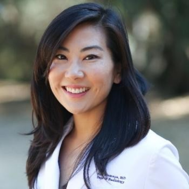
Aya Kamaya, M.D.
Professor of Radiology
Division Chief
Edward Lo, M.D.
Clinical Assistant Professor
Nayeli Morimoto, M.D.
Clinical Associate Professor
Lindsey Negrete, M.D.
Clinical Assistant Professor
Richa Patel, MD
Clinical Instructor
Peter Poullos, M.D.
Clinical Associate Professor
Luyao Shen, M.D.
Clinical Assistant Professor
Andrew Shon, M.D.
Clinical Assistant Professor
Justin Tse, M.D.
Assistant Professor
Volney Van Dalsem, M.D.
Clinical Professor
Luke Yoon, M.D.
Clinical Associate Professor
Conferences & Teaching
- Body Rounds & Journal Club: This weekly CME-accredited conference is attended by all Body Imaging Fellows, residents, and body imaging faculty. Interesting cases from the week are presented and discussed. Journal Club is held monthly, with in-depth discussion of the selected journal club article, followed by presentation of clinical cases pertaining to the article.
- Weekly Case Conference: This is an informal conference that is attended by faculty, fellows, and residents. Interesting cases are routinely shown during this conference, as well as difficult cases that stimulate discussions and opinions from the participants. These conferences are intermixed with peer review, prostate tumor board, interesting cases on body MRI.
- Tumor Boards: Body Imaging Fellows routinely present cases at interdisciplinary tumor boards such as Hepatobiliary, Neuroendocrine, and Colorectal conferences, which provides a rich environment of teaching and exposure to interesting pathology.
- Grand Rounds: Twice a month at this CME-accredited conference, invited guests from around the world, as well as Stanford faculty, present lectures on various topics.
- Ultrasound Scanning Club: Once a week, Body Fellows focus on hands-on scanning of a specific anatomic structure with dedicated instruction on proper scanning technique, image optimization, and tips and trick in image acquisition.
- Didactic Fellow Lectures: This lecture series runs during July through September and is specifically designed for the Body Imaging Fellows, with presentations by the body imaging, cardiovascular, and MRI faculty. Topics covered include:
Acute abdominal vascular disorders (J. Shen)
Acute aorta I (Fleischmann)
Acute aorta II (Fleischmann)
Acute pulmonary embolism (Becker)
Appendicitis (Jeffrey)
Biopsy workshop (Negrete & L. Shen)
BMT complications (Desser)
Cardiac assist devices (J. Shen)
Chest trauma & X-ray of foreign bodies (Leung)
Congenital internal hernias and volvulus (Chow)
CT colonography (Poullos)
Emergency body MR - protocols and interpretation (Syed)
Endometriosis (Jha)
ER body imaging pitfalls (Hsu)
First trimester US (Kamaya)
IV and oral contrast, contrast reactions, and indications (Morimoto)
Liver doppler (Kamaya)
Liver THIDS & THADS (Desser)
Liver transplant evaluation (Kamaya)
Mesenteric Ischemia (Chow)
MR defecography (Sheth)
MR of cholangiocarcinoma (Brunsing)
O-RADS US and MRI (Jha)
Pelvic floor MRI (Sheth)
Perianal Fistula (Syed)
Plumbing 101: post surgical anatomy and complications (Poullos)
Post partum complications (Kamaya)
Prostate MRI (Loening)
Protocoling CT (J. Shen)
Quantitative cancer imaging & RECIST (Becker)
Right upper quadrant pain (Kamaya)
Rectal cancer staging MRI (Sheth)
Thyroid imaging and TI-RADS (Kamaya)
Inflammatory bowel disease (Patel)
Online Medical Education Lectures by Body Imaging Faculty
Terry Desser, MD:
Thyroid Nodules: What We Know and What We Don't
R. Brooke Jeffrey, MD:
Complicated Cholecystitis
Ultrasound for RLQ Pain: What's New?
Ultrasound of Cervical Lymph Nodes
Aya Kamaya, MD:
HCC Surveillance And Fibrosis MR
Ultrasound Screening of Hepatocellular Carcinoma - Part 1
Color Doppler Sonography in Genitourinary Ultrasound
Principles, Concepts, and Applications of Ultrasonography
SRU Gallbladder Polyp Consensus Committee Guidelines
Multicenter Study of ACR Ultrasound LI-RADS Visualization Scores on Serial Examinations
Longitudinal Ultrasound Assessment of Changes in Size and Number of Gallbladder Polyps
Lindsey Negrete, MD:
Let's Read Out Podcast
Peter Poullos, MD:
Thriving after life-changing trauma
Justin Tse, MD:
Growth Kinetics and Progression Rate of Bosniak Classification, Version 2019 III and IV
Bosniak Classification of Cystic Renal Masses: Comparison of Categorization using CT & MRI