Quantitative MRI of Regional Renal Function
By accurately calculating the glomerular filtration rate (GFR) with MRI, we can better assess kidney health and diagnose chronic kidney disease. Healthy kidneys filter the blood to remove waste, but this function can be impaired in diseased kidneys, leading to a decreased GFR. In dynamic MR urography, a Gadolinium-based contrast agent is administered intravenously and a series of images is acquired to track the uptake and clearance of this contrast agent in the kidneys. A pharmokinetic model can be used to analyze the contrast exchange between the arteries and the kidneys and estimate the GFR. In order to get accurate GFR maps, we need high temporal resolution for the arteries, high spatial resolution for the kidneys, and minimal respiratory motion artifacts. In this work, we developed a method to enable free-breathing scans with high spatial and temporal resolution, and improved GFR accuracy by performing an additional reconstruction to better estimate the concentration-time curves in a large arterial region of interest. Currently there is no method to measure the GFR in different regions, so this approach has the potential to help diagnose and locate problems with kidney function.
Yoruk U, Saranathan M, Loening AM, Hargreaves BA, Vasanawala SS. High temporal resolution dynamic MRI and arterial input function for assessment of GFR in pediatric subjects. Magn Reson Med. 2016 Mar;75(3):1301-11.
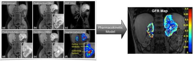
Quantitative maps of glomerular filtration rate (GFR) are useful for diagnosing chronic kidney disease.
Umit Yoruk and Manoj Saranathan are alumni of the BMR group