A Simple Analytic Method for Estimating T2 in the Knee from DESS
MRI measurements of the relaxation time T2 can help estimate the progression of diseases such as osteoarthritis and its associated tissue deterioration. The Double-Echo Steady-State (DESS) sequence is often used for radiographic images of the knee. The sequence produces two signals, S1 and S2. The ratio S2/S1 can be used to compute the T2 value of every pixel in the image. However, doing this accurately is complicated and requires time-consuming numerical computational methods or inaccurate approximations. In this work, we developed a technique that provides a simple yet accurate estimation of T2 from a single DESS scan, typically 3-5min. The technique involves a new model for the signal ratio S2/S1, which is highly accurate under a variety of conditions and much faster than standard numerical computations.
Sveinsson B, Chaudhari AS, Gold GE, Hargreaves BA. A Simple Analytic Method for Estimating T2 in the Knee from DESS. Magn Reson Imaging. 2017 May;38:63-70.
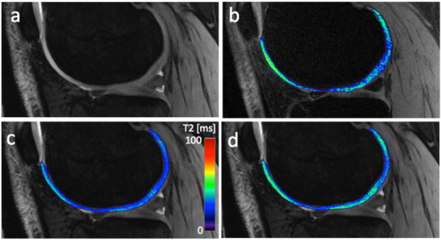
(a) An S1 image from DESS without any T2 map. (b) A T2 map in the femoral cartilage acquired with FSE, a standard, slower sequence. (c) A T2 map obtained by an older, fast processing method of DESS. The map shows lower values than the FSE map. (d) A T2 map obtained with the proposed, fast processing method. The map is very similar to the FSE map.
Bragi Sveinsson is an alumnus of the BMR group