Rapid 3D MRI of Breast Perfusion
Breast cancer is most often detected on MRI using contrast-enhanced imaging. Following injection of gadolinium, the signal brightness increases at different rates as the agent reaches different tissues, and tumors enhance more quickly. There is a tradeoff between how quickly we can image (temporal resolution), and image sharpness. Our proposed method allows a favorable tradeoff, and also allows the temporal resolution to be varied throughout the scan, so that sharper images are acquired during periods of slower signal change. This is now the routine approach for breast MRI at many centers including Stanford.
Saranathan M, Rettman DW, Hargreaves BA, Lipson J, Daniel BL. Variable spatiotemporal resolution three-dimensional Dixon sequence for rapid dynamic contrast-enhanced breast MRI. J Magn Reson Imaging. 2014 Dec;40(6):1392-9.
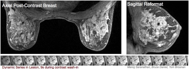
Axial and sagittal (side) slices from a 3D dynamic contrast-endhanced breast MRI acquisition. Increased and leaky vasculature to tumors results in rapid uptake of contrast, and the tumor is bright on these images.
Manoj Saranathan is an alumnus of the BMR group