2D Multi-Spectral Imaging For Fast MRI Near Metal
This approach offers a fast (tens of seconds per slice) approach to MRI near metal while correcting much of the signal loss, distortion and abnormally bright signal that can occur near implanted metal.
While many passive implanted metal devices are safe for MRI scanning, they cause severe magnetic field variations that cause artifacts. Reversing the sign of the slice selection gradient in common spin-echo methods can enable selective excitation of both limited slice and limited frequency range. This allows separate imaging of the wide range of signal frequency bands that would otherwise result in distortion and other artifacts. The signal for a slice is built up by different frequency bands, which are combined to form a final image.
Hargreaves BA, Taviani V, Litwiller DV, Yoon D. 2D multi-spectral imaging for fast MRI near metal. Magn Reson Med. 2018 Feb;79(2):968-73.
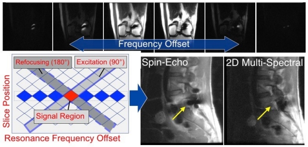
Each slice is separated into individual frequency bands that are imaged separately by reversing the slice-selection gradient in a spin-echo method. The end result is a fast approach (12seconds per slice here) that reduces the artifacts and in this case allows visualization of nerve roots in the spine that are obscured in standard spin-echo images (arrows).

Valentina Taviani is an alumnus of the BMR group