Bhutani Lab Research Program
Our multidisciplinary team is broadly interested in understanding cellular mechanisms that operate in skeletal tissues in health, aging, and disease. We are driven towards understanding biology and in the future translate these fundamental discoveries towards unmet medical needs in the clinic.
High-Dimensional Single Cell Omics for Skeletal Tissues
New cutting-edge molecular biology tools have the power to provide a high resolution understanding of single cells and their variability instead of a snapshot of the total population of cells - such an understanding is invaluable to understand both ‘protective’ (stem and progenitor cells) as well as disease-propagating rare cell populations (cancer stem cells, senescent cells, Inflammation-amplifying cells).
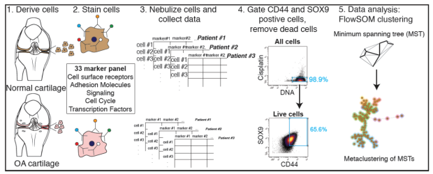
To accomplish this, we utilize multi-omics methods-single cell RNA sequencing, sc-ATAC seq, CyTOF for isolated cells and CODEX (CO-Detection by indEXing) on fixed tissues-thereby building transcriptomic, epigenomic and proteomic cellular atlases for skeletal tissues (including cartilage, bone, synovium). Mouse models of disease, transgenic mice and patient samples (OA and cancer) are used in parallel to get a comprehensive and clinically relevant insights into the cell populations and neighborhoods that operate in skeletal health and disease.
The Minority Report - Understanding and Harnessing Patient Heterogeneity for Precision Medicine
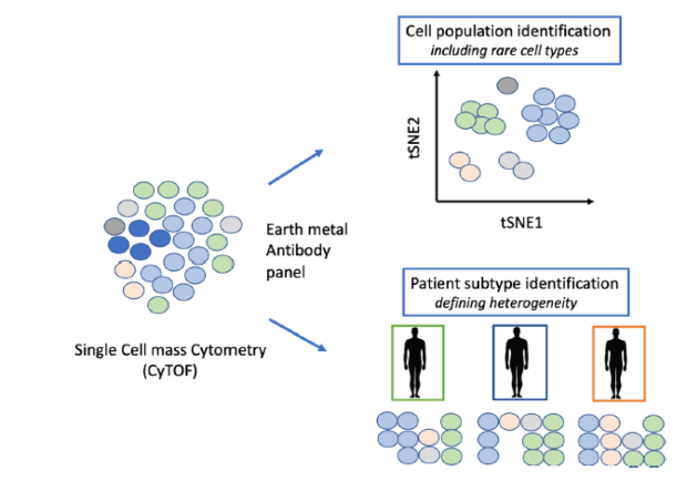
Another research interest is to utilize single cell approaches (CyTOF and CODEX) to increase the understanding of patient heterogeneity - in OA and skeletal cancers. By utilizing cohorts of healthy donors and patients, we are able to build a landscape of the distinct cell populations that exist in disease relevant tissues and immune cell populations.
Mapping this landscape now provides the tools to test how these populations respond to putative drugs and how uniform or variable this response is within a cohort of patients. We aim to extend this approach to a much larger cohort of OA patients to identify subsets of patients that could respond to a particular drug thereby making possible a ‘precision medicine’ approach to find the right drug for the right patient.
Epigenetic Regulation in Development and Disease
DNA methylation is an epigenetic mark associated with long-term gene silencing during early development and lineage specification. Studies by our group and others uncovered novel DNA repair based DNA demethylation pathways and new cytosine modifications (5hmC, 5caC and 5fC). The role and effect of 5hmC on 5mC turnover and gene expression is poorly understood. We are exploring the role of these novel DNA demethylation regulators in cartilage development, regeneration and disease.
Understanding Osteoarthritis Pathogenesis for Novel Therapeutics
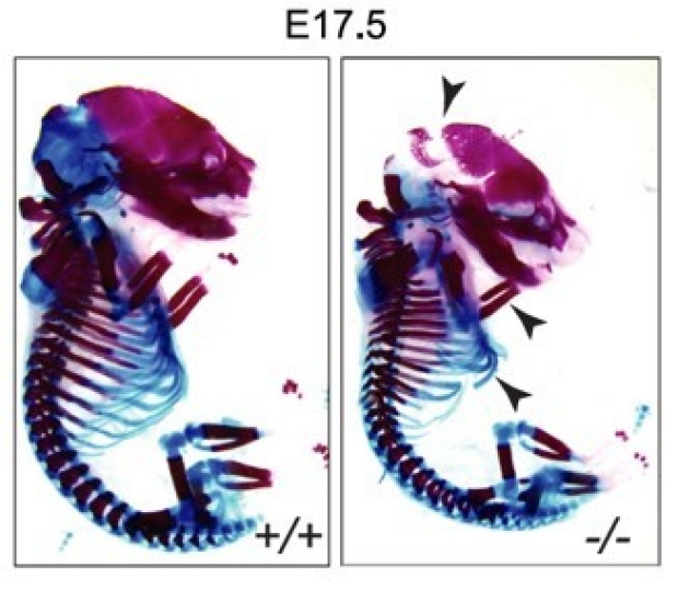
Osteoarthritis (OA) is an age-associated multifactorial disease characterized by joint dysfunction and cartilage degeneration that affects as much as 40% of the elderly population. Despite OA causing a medical deficit of billions of dollars per year, it remains an unmet medical need with not a single disease-modifying drug on market. In this research program, we postulated that epigenetic aberrations could play a major role in OA pathology akin to cancer. Using global sequencing technologies, we identified that epigenetic patterns in DNA hydroxymethylation (5hmC) are greatly altered in cartilage from OA patients and contribute to the aberrant disease-specific gene expression.
Interestingly, upon utilizing (a) transgenic mice that are deficient in these epigenetic modifications and (b) specific epigenetic inhibitors in normal mice, we observe that OA in a surgical mice model can be attenuated. This putative small molecule therapeutic can protect the mice as well as human end-stage osteoarthritic cartilage. Ongoing and future studies are aimed to understand the ‘early’ and ‘late’ stages of OA pathogenesis and the potential of these inhibitors to reverse. Overall, this research direction aims to evaluate a potential new therapeutic axis for OA based on epigenetic modulation, with newer explorations in higher chromatin organization and long noncoding RNA.
Engineering Induced Pluripotent Stem Cells (iPSC) Based Cells, Tissues, and Organoids
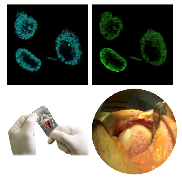
The discovery of induced pluripotency by Yamanaka and colleagues has revolutionized the field of regenerative medicine. Induced pluripotent stem cells (iPSC), generated by introduction of a few defined factors in a somatic cell, provide an ideal patient-specific source for disease modeling, drug discovery and cellular therapies. Our research is geared towards applying reprogramming approaches towards musculoskeletal regeneration especially cartilage regeneration that remains an unmet medical need. We have optimized quick and efficient differentiation methodologies to generate chondrocytes and mesenchymal stromal cells from human iPSCs. These human iPSC-derived cells are now being utilized to address the molecular mechanisms of cartilage aging and regeneration, including novel factors that differentiate young and old cartilage. In addition, these reprogramming and differentiation tools are being used for organoid generation, modeling pediatric growth disorders and for drug screening using small molecule libraries.
Unlocking the Fountain of Youth - Defining Cellular Senescence and Opposing Juvenile Factors

Human cartilage regeneration is inherently limited; hence cartilage injuries persist even in young and healthy adults, increasingly translated to early onset of Osteoarthritis. Clinically, it has been long known that cartilage repair is more effective in pediatric populations. Based on this premise, we have utilized multiple global gene and protein expression analyses to identify a group of ‘novel’ juvenile factors that are lost with age. These factors show a unique potential in rejuvenation of joint tissues and we are now testing how to harness their ability to benefit aged and diseased joints.
Biomimetic Scaffolds and Microfluidics Approaches for Tissue Engineering
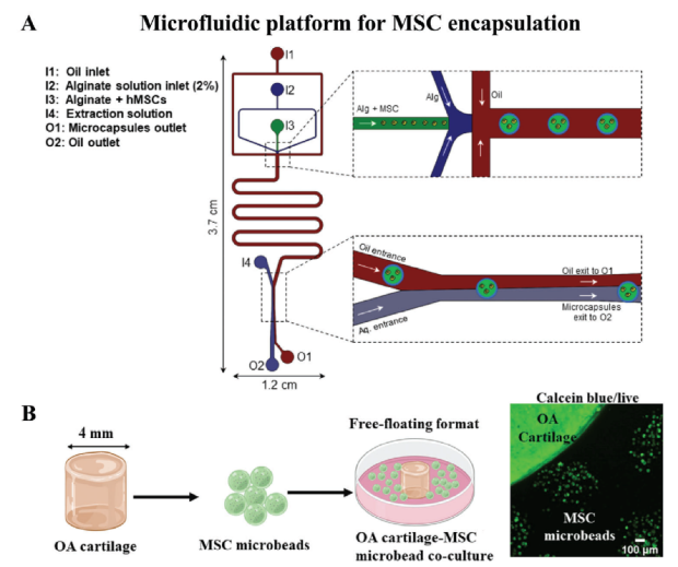
Finally, we are applying multiple bioengineering principles towards optimal tissue engineering including microfluids platforms and hydrogel based scaffolds with varying biophysical and mechanical properties.
One area of interest is optimizing scaffolds for engineering autologous cartilage tissue from iPSC generated from human patient samples and testing its potential for repairing cartilage defects in animal models of focal cartilage repair. Secondly, we have developed new encapsulation processes for increased survival and function of mesenchymal stromal cells that enhances their ability to immunomodulate a given microenvironment.

We are also investigating the fundamental interaction between the cell and its pericellular or extracellular matrix to understand how the external ‘biophysical’ cues are sensed and transduced by the cell. Our recent studies have identified that a mechanosensitive surface calcium channel Trpv4 and GSK as the signaling pathway that allows a chondrocyte to sense changes in ECM viscoelasticity.
Changes in ECM viscoelasticity alone were sufficient to induce cell volume restriction and inflammatory pathways in the cell, dramatically altering its cell state. In addition, we discovered that this pathway was dysfunctional in diseased chondrocytes suggesting an intricate interplay between the ECM characteristics and cell fate that likely plays a significant role in development and disease.